Patellar Tendinitis: Causes and Symptoms

Article featured on Mayo Clinic

Overview

Patellar tendinitis is an injury to the tendon connecting your kneecap (patella) to your shinbone. The patellar tendon works with the muscles at the front of your thigh to extend your knee so that you can kick, run and jump.

Patellar tendinitis, also known as jumper’s knee, is most common in athletes whose sports involve frequent jumping — such as basketball and volleyball. However, even people who don’t participate in jumping sports can get patellar tendinitis.

For most people, treatment of patellar tendinitis begins with physical therapy to stretch and strengthen the muscles around the knee.

Symptoms

Pain is the first symptom of patellar tendinitis, usually between your kneecap and where the tendon attaches to your shinbone (tibia).

Initially, you may only feel pain in your knee as you begin physical activity or just after an intense workout. Over time, the pain worsens and starts to interfere with playing your sport. Eventually, the pain interferes with daily movements such as climbing stairs or rising from a chair.

When to see a doctor

For knee pain, try self-care measures first, such as icing the area and temporarily reducing or avoiding activities that trigger your symptoms.

Call your doctor if your pain:

  • Continues or worsens
  • Interferes with your ability to perform routine daily activities
  • Is associated with swelling or redness about the joint

Causes

Patellar tendinitis is a common overuse injury, caused by repeated stress on your patellar tendon. The stress results in tiny tears in the tendon, which your body attempts to repair.

But as the tears in the tendon multiply, they cause pain from inflammation and weakening of the tendon. When this tendon damage persists for more than a few weeks, it’s called tendinopathy.

Risk factors

A combination of factors may contribute to the development of patellar tendinitis, including:

  • Physical activity. Running and jumping are most commonly associated with patellar tendinitis. Sudden increases in how hard or how often you engage in the activity also add stress to the tendon, as can changing your running shoes.
  • Tight leg muscles. Tight thigh muscles (quadriceps) and hamstrings, which run up the back of your thighs, can increase strain on your patellar tendon.
  • Muscular imbalance. If some muscles in your legs are much stronger than others, the stronger muscles could pull harder on your patellar tendon. This uneven pull could cause tendinitis.
  • Chronic illness. Some illnesses disrupt blood flow to the knee, which weakens the tendon. Examples include kidney failure, autoimmune diseases such as lupus or rheumatoid arthritis and metabolic diseases such as diabetes.

Complications

If you try to work through your pain, ignoring your body’s warning signs, you could cause increasingly larger tears in the patellar tendon. Knee pain and reduced function can persist if you don’t tend to the problem, and you may progress to the more serious patellar tendinopathy.

Prevention

To reduce your risk of developing patellar tendinitis, take these steps:

  • Don’t play through pain. As soon as you notice exercise-related knee pain, ice the area and rest. Until your knee is pain-free, avoid activities that put stress on your patellar tendon.
  • Strengthen your muscles. Strong thigh muscles are better able to handle the stresses that can cause patellar tendinitis. Eccentric exercises, which involve lowering your leg very slowly after extending your knee, are particularly helpful.
  • Improve your technique. To be sure you’re using your body correctly, consider taking lessons or getting professional instructions when starting a new sport or using exercise equipment.

The Orthopedic & Sports Medicine Center of Oregon is an award-winning, board-certified orthopedic group located in downtown Portland Oregon. We utilize both surgical and nonsurgical means to treat musculoskeletal trauma, spine diseases, sports injuries, degenerative diseases, infections, tumors and congenital disorders.

Our mission is to return our patients back to pain-free mobility and full strength as quickly and painlessly as possible using both surgical and non-surgical orthopedic procedures.

Our expert physicians provide leading-edge, comprehensive care in the diagnosis and treatment of orthopedic conditions, including total joint replacement and sports medicine. We apply the latest state-of-the-art techniques in order to return our patients to their active lifestyle.

If you’re looking for compassionate, expert orthopedic surgeons in Portland Oregon, contact OSM today.

Phone:
503-224-8399

Address
1515 NW 18th Ave, 3rd Floor
Portland, OR 97209

Hours
Monday–Friday

Knee Hyperextension: What is it?

Article featured on News Medical Life Sciences

Knee hyperextension is a condition caused when the knee straightens too far, beyond the normal maximum limit of 00 and often with the joint in varus malalignment. In the properly aligned knee, the load is borne on a line running down the center of the hip, knee and ankle, but in a varus deformity (bowleg form), the line is shifted outwards and back.

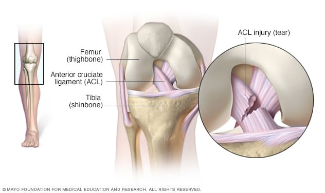
The most common injuries are posterolateral joint injury and avulsion of the bone attached to the anterior cruciate ligament, and they are especially common in young children due to their softer bones. Posterior cruciate ligament, however, is rare.

Causes

Knee hyperextension may occur because of:

  • Trauma caused by impact to the front of the knee, which makes the joint move backward, putting high strain on the anterior cruciate ligament – such as when a football player faces a leg tackle.
  • Pushing the femur or patella over the tibia, the lower leg bone, for example when one slides to a sudden stop using one leg, which stresses the major ligaments within the knee.
  • Genu recurvatum: this is a deformity characterized by knee hyperextension over 5 degrees.
  • Nerve damage, such as Brain injury (congenital, stroke-induced or traumatic), or poliomyelitis, may cause hyperextended gait because of muscle (quadriceps or calf muscle) atrophy, spastic plantar flexion of the ankle, or contractures of the heel tendons.

Symptoms

The cause of hyperextension predicts the symptoms, which may be minor pain and swelling when it occurs to a trivial degree, but may consist of sharp pain, strained or torn cruciate ligaments, avulsion of bone chips accompanying such tears, and bone bruising may occur on the anterior part of the knee joint. When the injury is at the posterolateral aspect, the ACL and the PCL typically tears. The rate of strain doesn’t seem to be the major factor as anterior cruciate ligaments tears vary at all rates of strain. Genu recurvatum may present with knee pain, abnormal gait, and a lack of proprioceptive perception, which makes it difficult to tell when the terminal extension of the knee is attained.

The hyperextended knee gait is marked by various degrees of abnormality. In some, the patient can walk only with the aid of a cane or a crutch. In others with minimal weakness, the knee becomes hyperextended only when the patient walks too much or indulges in other heavy work or exercise, leading to muscle fatigue and loss of joint support. The associated ligamentary and muscle weakness and joint integrity also contribute to the final abnormality of gait, as does the presence of arthritis in the knee joint.

Another symptom is knee instability, or a feeling of giving way at the knee, in part or full, during normal activities.

Pain may be felt over the knee or to the medial side, and is caused by compression of the soft tissues by the malaligned knee, as well as on the posterolateral aspect, due to stretching of the soft tissue. In acute hyperextension injury the knee may pop and acute swelling often occurs within a few hours of the injury. The pain can become too severe for normal support of weight during walking or running.

Anatomy

The knee joint is supported on the lateral and posterior aspects by the fibular collateral ligament, and the popliteus muscle with its tendon and ligament. This complex of structures keeps the knee compartment from widening laterally, prevents dislocation of the lateral surface of the tibial component of the knee to the back, keeps the tibia from rotating, and thus prevents both knee hyperextension and genu recurvatum. The bones may show varus alignment, in some cases. A triple varus knee is caused by three factors:

  • Tibiofemoral alignment is disturbed.
  • The lateral tibiofemoral compartment separation is increased because of weak supporting structures on the posterolateral aspect of the knee.
  • The knee appears bowlegged in skeletal outline when fully extended.

In most cases both the posterolateral structures and the anterior cruciate ligament is damaged or at least weak, as following a knee injury or any other cause of muscle atrophy. Patellofemoral arthritis is another cause, but in this case the hyperextended knee gait is due to pain as well as muscle weakness.

Diagnosis

The history will offer clues to the diagnosis, such as prior knee injury. A physical examination will help confirm and grade the injury, including joint mobility, visible injury, bruising or swelling, and locking of the joint. Imaging is needed in severe injury and especially if surgery is contemplated. MRI and X-ray imaging are typically performed.

Treatment

Minor hyperextension of the knee may require only the R.I.C.E approach:

  • Rest and avoidance of physical activities that strain the knee in any way for a few weeks.
  • Ice application several times a day.
  • Compression using a knee brace and crutches for support are helpful in protecting the ligaments of the knee against any further damage.
  • Elevation to reduce edema.

Severe cases will require physical therapy, with graded exercises of the quadriceps and other hip and knee muscles. This should be done under supervision so that further injury does not occur, and full joint mobility is attained. Surgery is required to reattach torn ligaments and will again be followed by physical therapy.

Correction of the hyperextended knee gait is crucial if the deformity is to be corrected permanently, otherwise the excessive tensile force on the ligaments inside the joint and increased muscle force could increase the load on the joint capsules, especially the medial and lateral compartments. This can be harmful to joint integrity in varus malalignment.


The Orthopedic & Sports Medicine Center of Oregon is an award-winning, board-certified orthopedic group located in downtown Portland Oregon. We utilize both surgical and nonsurgical means to treat musculoskeletal trauma, spine diseases, sports injuries, degenerative diseases, infections, tumors and congenital disorders.

Our mission is to return our patients back to pain-free mobility and full strength as quickly and painlessly as possible using both surgical and non-surgical orthopedic procedures.

Our expert physicians provide leading-edge, comprehensive care in the diagnosis and treatment of orthopedic conditions, including total joint replacement and sports medicine. We apply the latest state-of-the-art techniques in order to return our patients to their active lifestyle.

If you’re looking for compassionate, expert orthopedic surgeons in Portland Oregon, contact OSM today.

Phone:
503-224-8399

Address
1515 NW 18th Ave, 3rd Floor
Portland, OR 97209

Hours
Monday–Friday

Swollen Knee Causes and Treatments

Article featured on the Mayo Clinic

Overview

A swollen knee occurs when excess fluid accumulates in or around your knee joint. Your doctor might refer to this condition as an effusion (ih-FYU-zhen) in your knee joint. Some people call this condition “water on the knee.”

A swollen knee may be the result of trauma, overuse injuries, or an underlying disease or condition. To determine the cause of the swelling, your doctor might need to obtain a sample of the fluid to test for infection, disease or injury.

Removing some of the fluid also helps reduce the pain and stiffness associated with the swelling. Once your doctor determines the underlying cause of your swollen knee, appropriate treatment can begin.

Symptoms

Signs and symptoms typically include:

  • Swelling. The skin around your kneecap can puff up noticeably, especially when you compare the affected knee to the normal one.
  • Stiffness. When your knee joint contains excess fluid, you might not be able to bend or straighten your leg completely.
  • Pain. Depending on the cause of the fluid buildup, the knee might be very painful — to the point that it’s difficult or impossible to bear weight on it.

When to see a doctor

See your doctor if:

  • Self-care measures or prescribed medications don’t relieve the pain and swelling
  • One knee becomes red and feels warm to the touch compared to your other knee

Causes

Many types of problems, ranging from traumatic injuries to diseases and other conditions, can cause a swollen knee.

Injuries

Damage to any part of your knee can cause excess joint fluid to accumulate. Injuries that can cause fluid buildup in and around the knee joint include:

  • Torn ligament, particularly the anterior cruciate ligament (ACL)
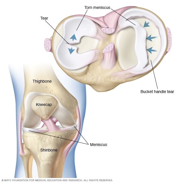
  • Cartilage (meniscus) tear
  • Irritation from overuse
  • Broken bones

Diseases and conditions

Underlying diseases and conditions that can produce fluid buildup in and around the knee joint include:

  • Osteoarthritis
  • Rheumatoid arthritis
  • Infection
  • Gout
  • Pseudogout
  • Bursitis
  • Cysts
  • Tumors

Risk factors

  • Age. Your likelihood of developing a swollen knee related to arthritis increases as you age.
  • Sports. People who participate in sports that involve twisting the knee, such as basketball, are more likely to experience the types of knee injuries that cause swelling.
  • Obesity. Excess weight puts added stress on the knee joint, contributing to the tissue and joint overload and knee degeneration that can lead to a swollen knee. Obesity increases your risk of osteoarthritis, one of the more frequent causes of knee swelling.

Complications

Complications of a swollen knee can include:

  • Muscle loss. Fluid in the knee can harm the working of your muscles and cause thigh muscles to weaken and atrophy.
  • Fluid-filled sac (Baker’s cyst). The buildup of fluid in your knee can lead to the formation of a Baker’s cyst in the back of your knee. A swollen Baker’s cyst can be painful, but usually improves with icing and compression. If the swelling is severe, you might need to have fluid removed (cyst aspiration).

Prevention

A swollen knee is typically the result of an injury or chronic health condition. To manage your overall health and prevent injuries:

  • Strengthen the muscles around your knee. Strong muscles around a joint can help ease pressure on the joint itself.
  • Choose low-impact exercise. Certain activities, such as water aerobics and swimming, don’t place continuous weight-bearing stress on your knee joints.
  • Maintain a healthy weight. Excess weight contributes to the wear-and-tear damage that can lead to a swollen knee.

The Orthopedic & Sports Medicine Center of Oregon is an award-winning, board-certified orthopedic group located in downtown Portland Oregon. We utilize both surgical and nonsurgical means to treat musculoskeletal trauma, spine diseases, sports injuries, degenerative diseases, infections, tumors and congenital disorders.

Our mission is to return our patients back to pain-free mobility and full strength as quickly and painlessly as possible using both surgical and non-surgical orthopedic procedures.

Our expert physicians provide leading-edge, comprehensive care in the diagnosis and treatment of orthopedic conditions, including total joint replacement and sports medicine. We apply the latest state-of-the-art techniques in order to return our patients to their active lifestyle.

If you’re looking for compassionate, expert orthopedic surgeons in Portland Oregon, contact OSM today.

Phone:
503-224-8399

Address
1515 NW 18th Ave, 3rd Floor
Portland, OR 97209

Hours
Monday–Friday
8:00am – 4:30pm

Why Do My Knees Pop?

Article featured on Cone Health Medical Group

Have you ever bent down to pick up something and heard a loud pop or crack coming from your knees? This sound is called “crepitus,” which is defined as “joint noise.” Popping knees are not unusual. It happens when carbon dioxide builds up in the joint’s synovial fluid and is released as a gas bubble that bursts when the joint adjusts rapidly. It is the same process that causes knuckles to crack.

Most of the time, this noise should cause no concern. There has been a rumor circulating for years that popping joints are a sign of impending arthritis, but there has been no research that supports this.

Some folks may hear a grinding noise in the knee when they squat. This is another form of crepitus and is typically nothing to be concerned about. The sound is caused by the cartilage rubbing on the joint surface and other soft tissue when the knee moves.

Most people experience crepitus their entire lives with no problems.

What if it hurts when my joints pop or grind?

You should be concerned if you have joint noise that is accompanied by pain, discomfort or swelling. This could be a sign that medical attention is required. Two of the most common cause for knee pain are:

  1. Cartilage injury or wear. The underside of your kneecap can unevenly rub on the front of your femur (thigh bone) and cause the cartilage under your kneecap to soften and wear off. Once the cartilage wears off, it hangs around the joint and can irritate the surrounding area. This irritation can result in pain when squatting, climbing stairs or even sitting for an extended period.
  2. Meniscus tear. The rubbery disc that cushions your knee and absorbs shock is called the meniscus. It can tear when there is an unexpected twist or turn in the joint, usually when the knee moves and the foot stays stationary. With aging comes greater risk of a meniscus tear, even through everyday activities.

What are the treatment options for knee injuries?

  • Rest – Stay off your knee as much as possible.
  • R.I.C.E. – Rest, ice, compression and elevation will promote healing and reduce swelling.
  • Knee Brace – There are many kinds, so it is best to consult your care provider for advice.
  • Physical therapy – A physical therapist can help you learn techniques and exercises to strengthen your knee and allow it time to heal.
  • Surgery – Minimally invasive surgery, especially for meniscus tears, can repair the tear and clear out any tissue causing pain, swelling and irritation.  

To help protect the knees, try exercises that develop the quadriceps, which are the muscles in the front of the thigh. Exercises that benefit this muscle group are walking, cycling and swimming. Other effective ways of protecting the knees are wearing supportive shoes, warming up before and stretching after exercise, and maintaining a healthy body weight.


The Orthopedic & Sports Medicine Center of Oregon is an award-winning, board-certified orthopedic group located in downtown Portland Oregon. We utilize both surgical and nonsurgical means to treat musculoskeletal trauma, spine diseases, sports injuries, degenerative diseases, infections, tumors and congenital disorders.

Our mission is to return our patients back to pain-free mobility and full strength as quickly and painlessly as possible using both surgical and non-surgical orthopedic procedures.

Our expert physicians provide leading-edge, comprehensive care in the diagnosis and treatment of orthopedic conditions, including total joint replacement and sports medicine. We apply the latest state-of-the-art techniques in order to return our patients to their active lifestyle.

If you’re looking for compassionate, expert orthopedic surgeons in Portland Oregon, contact OSM today.

Phone:
503-224-8399

Address
1515 NW 18th Ave, 3rd Floor
Portland, OR 97209

Hours
Monday–Friday
8:00am – 4:30pm
Did You Know That Inactivity is Actually HARD On Your Knees?

Between working from home and being glued to the television watching the latest election updates, many of us are guilty of increased “couch potato” behavior lately.  And we’ve all heard of the dangers of sedentary lifestyles contributing to obesity, etc.,  but did you know that *not* moving can actually weaken your knees and increase your chances of osteoporosis?

Continue reading for more from Noyes Knee Institute and the Journal of Public Health.

Do you spend a lot of time sitting? Maybe you work at an office where most of your time is at your desk, or maybe when you’re at home, you prefer to rest on the couch instead of being on your feet. Many people live a mostly inactive lifestyle, but they might not realize that inactivity can be the reason why they experience increased joint pain.

Learn the reasons why inactivity can hurt your knees and what you can do to change it.

Weakens Your Knees

If you live a life or limited activity, your body adapts to that lack of motion. Essentially, when you aren’t using your legs muscles, ligaments, and joints for moderate levels of activity, you are losing them. Your knees become weaker as you require less of them.

One runner found that as she took time off running to rehabilitate an injury, she could not run after completing her recovery because of knee pain. She had to complete additional physical therapy because the rest had caused her to develop a condition called chondromalacia of the patella.

Essentially, her kneecap would not follow the proper range of motion because she had developed some weakness in the joint. It’s a common condition for people who are not active. Even something as simple as going up or down the stairs can make your knees ache.

If you spend your day sitting, you also experience pain in other areas that can also aggravate the knee. Your quadriceps become tight, which exert a pulling sensation on your knees.

You can help your knees feel better by focusing on flexibility. Stretch daily, and participate in joint-stabilizing exercises like yoga. Try to be more active during the day. Stand at your desk, or take time to walk around the office a few times. Use the stairs instead of the elevator.

Increases Your Risk of Arthritis Pain

If you start to experience joint pain as a result of arthritis, your first instinct is to rest more, because walking, running, biking, or other activities cause pain. However, resting when you have knee pain is often the worst solution.

Resting will cause the muscles that give the knee support to weaken. As a result, they become less able to bear your weight, which results in greater joint pain. Reduced strength in the knee joint also translates to reduced stability, which can increase your risk for accidents and make exercising even more difficult to do safely.

If you have arthritis or if you have a history of arthritis in your family, staying active is one way you can help to prevent it from getting worse. Ask a knee specialist for exercises that are safe and helpful for strengthening your knees without causing you too much pain during workouts.

Promotes Weight Gain

A sedentary lifestyle is often why people struggle to manage their weight. Gaining weight with age is common, and spending your days seated can make that problem worse. With every extra pound, the pressure on your knees increases by about four pounds. So, just 10 pounds of extra weight means 40 pounds of pressure on your knees.

All that stress naturally means that your knees start to hurt, and they can hurt even more when you try to be active again. Make sure you intentionally choose low impact exercise as first. Try a stationary bike or a brisk walk in supportive shoes to begin. Focus on losing weight through diet control.

After you lose some weight, you can increase your physical workouts if your doctor believes they will be safe. You might try incorporating some resistance training to really give your lower body some increased strength and stability, as long as you also spend time stretching and increasing your flexibility.

Increases Risk for Osteoporosis 

From the Daily Mail

Being a couch potato weakens your bones: Adults in their 60s face greater risk of fractures if they spend hours sitting down each day – but walking 10,000 steps each day helps

  • The study of 214 adults was published today in the Journal of Public Health
  • It is the first to show a link between a sedentary lifestyle and osteoporosis
  • Participants’ hips and spines were scanned to measure their bone density

A couch potato lifestyle leads to weaker bones in later life, particularly for men, researchers have found.

Experts discovered that men spent more time on average sitting still than women and therefore had weaker bones, particularly in their lower back.

But the new findings, conducted by academics from Durham and Newcastle universities, show that even just completing 10,000 steps a day can help to keep bones strong.

The study showed that people in their sixties who spent a lot of time sitting down had weaker bones which increased their risk of developing ‘fragility’ fractures.

It is well known that weight-bearing and muscle strengthening exercises are important for building bone strength and preventing osteoporosis.

The study, published in the Journal of Public Health, is the first to show that a sedentary lifestyle in men is associated with weaker bones and osteoporosis.

More than half a million fragility fractures – where a fracture occurs from a fall at standing height or less – happen each year in the UK. It is estimated that by 2025, that number will have gone up by 27 per cent.

Dr Karen Hind, of the Department of Sport and Exercise Sciences at Durham University, said: ‘We know that excessive sedentary time can lower someone’s metabolism which can lead to being overweight and Type 2 diabetes.

‘What we now know is that being inactive is also associated with lower bone strength and an increased risk of osteoporosis.

‘Osteoporosis is a disease that affects older people but by encouraging this age group to keep active, it will help improve their bone health.’

The research team followed 214 men and women, aged 62, from Newcastle University’s Thousand Families Study.

Each participant wore a monitor for seven consecutive days which measured their physical activity and sedentary time. The number of daily steps was also recorded, and then compared with public health recommendations.

The participants’ hips and spines were scanned to measure their bone density.

Participants involved in 150 minutes of light physical activity a week had better bone strength than the more sedentary participants, according to the findings.

The men who spent more than 84 minutes per day sitting still, compared to the average of 52 minutes, had 22 per cent lower bone density in their spine.

The researchers say the impact on their bone density is similar to that of smoking, which is also a risk factor for osteoporosis.

The economic and personal costs of osteoporosis are substantial – in the UK the direct costs of fragility fractures are estimated to be £4.4billion which includes £1.1billion for social care.

The participants all lived in Newcastle-upon-Tyne, and Public Health England statistics indicate that the North East has the greatest proportion of physically inactive adults and the highest incidence of hip fractures compared to the rest of the UK.

The researchers said that the message from their findings is: stay active and reduce sedentary time.

They emphasised that the study shows that hitting the daily target of 10,000 steps and avoiding long periods of sedentary time will increase bone strength.

They say that even making daily lifestyle ‘hacks’ can make a difference – such as parking the car further away from the shopping centre or taking the stairs instead of the lift.

Dr Hind added: ‘Currently there are no specific guidelines for this age group to encourage light physical activity or to reduce sedentary time.

‘Yet, as people retire they are more likely to increase the time they spend watching television and reduce their daily step count.

‘It would be great to see initiatives that specifically target this group to increase their awareness of the importance of staying active and reducing the amount of time spent sitting still.’

Learn more about bone health


The Orthopedic & Sports Medicine Center of Oregon is an award-winning, board-certified orthopedic group located in downtown Portland Oregon. We utilize both surgical and nonsurgical means to treat musculoskeletal trauma, spine diseases, sports injuries, degenerative diseases, infections, tumors and congenital disorders.

Our mission is to return our patients back to pain-free mobility and full strength as quickly and painlessly as possible using both surgical and non-surgical orthopedic procedures.

Our expert physicians provide leading-edge, comprehensive care in the diagnosis and treatment of orthopedic conditions, including total joint replacement and sports medicine. We apply the latest state-of-the-art techniques in order to return our patients to their active lifestyle.

If you’re looking for compassionate, expert orthopedic surgeons in Portland Oregon, contact OSM today.

Phone:
503-224-8399

Address
1515 NW 18th Ave, 3rd Floor
Portland, OR 97209

Hours
Monday–Friday
8:00am – 4:30pm
How can you tell if you have a torn meniscus?

How can you tell if you have a torn meniscus?

Article Featured on WebMD

Like a lot of knee injuries, a meniscus tear can be painful and debilitating. Unfortunately, it’s quite common. In fact, a meniscal tear is one of the most frequently occurring cartilage injuries of the knee. So how can you tell if you have one? Learn more in this article.
Read more
What is Compartment Syndrome?

What is Compartment Syndrome?

Article Featured on AAOS

Compartment syndrome is a painful condition that occurs when pressure within the muscles builds to dangerous levels. This pressure can decrease blood flow, which prevents nourishment and oxygen from reaching nerve and muscle cells.

Compartment syndrome can be either acute or chronic. Acute compartment syndrome is a medical emergency. It is usually caused by a severe injury. Without treatment, it can lead to permanent muscle damage.

Chronic compartment syndrome, also known as exertional compartment syndrome, is usually not a medical emergency. It is most often caused by athletic exertion.
Read more

Did I Tear my Meniscus? Symptoms of a Torn Meniscus

Did I Tear my Meniscus? Symptoms of a Torn Meniscus

Article Featured on Brigham Health

What is a torn meniscus?

There are 3 bones in the knee. These are the femur, tibia, and patella. The ends of those bones are covered with cartilage. This is a smooth material that cushions the bone and allows the joint to move easily without pain. The cartilage acts as a shock absorber. Between the bones of the knees are 2 crescent-shaped disks of connective tissue, called menisci. These also act as shock absorbers to cushion the lower part of the leg from the weight of the rest of the body.

Read more