What Does it Mean When Your Knee Pops?

Article featured on HSS

Decipher every snap, crackle and pop with this advice from a knee surgeon.

You’re getting up from your seat, walking across the room, or doing a weighted squat, and suddenly, a “pop” emanates from your knee. Should you be worried?

Not necessarily. “Most of the time, a popping sound is harmless,” says HSS orthopedic surgeon Dr. Sabrina Strickland. The official name for this noise is “crepitus” and there are several perfectly innocent reasons it can happen. Many times, the sound simply means air or gas bubbles have built up in the fluid around the joints and the cracking is those tiny bubbles bursting. Other times, knee crepitus can indicate early or even moderate osteoarthritis. “As the cartilage wears down, the underside of the kneecap rubs against the front of the thighbone, and a bend in the knee can cause a crackly, crunchy, creaky, or even pop-like sound,” says Dr. Strickland.

If you think you have osteoarthritis in your knee, you may want to consult a sports medicine physician or an orthopedic surgeon to make sure, especially if you are experiencing pain.

Injuries can also initiate knee pops. They include:

ACL tear

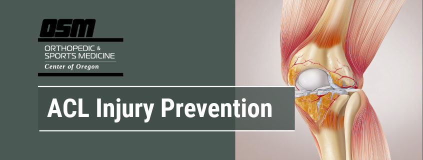
This is the most common injury associated with a pop. The anterior cruciate ligament (ACL) is one of four major ligaments in the knee joint. It helps maintain the knee’s rotational stability and prevents the tibia (shinbone) from slipping in front of the femur (thighbone). ACL tears, either partial or full, are especially common among people who play sports that require a lot of quick changes in direction, such as tennis, soccer, basketball and skiing. Active women are particularly at risk for tearing their ACL because their biomechanics tend to put more stress on their knees.

When a person tears their ACL, they usually hear or feel the pop at the time of the tear. The knee will quickly swell and often feel unstable. In less severe tears, the symptoms may be mild, while complete tears leave the person unable to land on the knee after a jump, accelerate then change directions, or pivot quickly.

While an ACL tear won’t heal on its own, not everyone needs surgery to repair it. “It really depends on which activities you want to get back to,” says Dr. Strickland. “You may get away without an ACL and still swim or do sports that work your knee in a straight line, like walking, jogging or cycling,” she says. But for activities that need a stable knee to twist or change direction, like skiing or tennis, an intact ACL is crucial.

Meniscus tear

The meniscus, a C-shaped piece of cartilage, spans the space between the thighbone and the shinbone. It acts as a shock absorber, keeping the bones of the knee joint from grinding against one other, and also helps transmit the load of your weight evenly across the knee joint.

A meniscus tear can happen while playing a sport where you twist your knee, like tennis or basketball, but it can sometimes tear when doing something as simple as a deep knee bend. Since less than the outer third of the meniscus contains blood vessels, the inner part never heals or regenerates. “Every step that you take, every time you go running, every time you jump off a step, you wear it down, so it’s very common to tear it without any significant trauma, particularly if you’re over 40,” says Dr. Strickland.

The key symptom of a meniscus tear is pain in the knee joint. A tear may also cause the knee to swell or bruise, and prompt a locking or catching sensation as the fragments of the meniscus gets caught inside the knee. The knee may also feel weak or give way for no apparent reason.

Unfortunately, because of the lack of blood supply, the meniscus can’t heal itself; however, not everyone needs surgery. Nonsurgical treatments, such as anti-inflammatory medications and rehabilitation with a physical therapist can help reduce the pain and strengthen and restore full motion of the knee. Other people will need surgery, which usually involves either a trimming or repair of the meniscus.

MCL tear

The medial collateral ligament (MCL) is located along the inner side of the knee, stretching from the thighbone to the shinbone and helps to stabilize the inner part of the knee. “MCL tears often happen while skiing, when the knee is subjected to a valgus force that causes the tibia to bend outwards relative to the femur,” says Dr. Strickland.

The pain is usually along the inner part of the knee, and there may be swelling and bruising. The good news is that MCL tears almost always heal on their own, and treatment consists of immediate rest, ice, compression, and elevation (RICE). For higher grade tears, patients benefit from use of a hinged knee brace to keep the ligament from stretching as it heals.

Baker’s cyst rupture

Many people don’t even know when they have one of these fluid-filled cysts that develop on the back of the knee. “Out of the blue, these harmless cysts can pop, causing the calf to swell and become discolored,” says Dr. Strickland. The pop is often accompanied by a sharp pain in the knee, and sometimes the feeling of fluid running down the calf. For most people, there’s no need for treatment, other than RICE. However, sometimes Baker’s cysts are due to problems with the knee joint, such as arthritis or a tear in the cartilage. “If that’s the case, we’ll have to treat the root cause,” says Dr. Strickland.

Patella subluxation

For most people, the kneecap, or patella, glides smoothly within a V-shaped groove in the thigh bone called the trochlea. As you bend and straighten the knee, the patella moves up and down and is stable within the trochlea, allowing you to stand, sit, walk, and run without a problem. But sometimes the kneecap partially goes off the trochlear groove, which is called patella subluxation, or can even fully dislocate. “When this happens, the knee swells, pops, or snaps,” says Dr. Strickland. This typically happens with younger people (under 25) who have a shallow groove.

Sometimes the kneecap can return to its proper place on its own. If not, a doctor can push it back in with gentle pressure, a process called reduction. Afterwards, rest, a knee brace, and crutches will help the swelling to go down and allow the knee to recover. Depending on the age of the patient, associated injuries, and radiologic measurements, some patients may require surgery to stabilize the patella.

The Orthopedic & Sports Medicine Center of Oregon

The Orthopedic & Sports Medicine Center of Oregon (OSM) is an award-winning, board-certified orthopedic and sports medicine practice serving Lake Oswego, Portland, Scappoose, and surrounding Oregon communities. Our main clinic is located in Lake Oswego, with additional locations in Portland and Scappoose.

OSM provides comprehensive orthopedic care, sports medicine, spine care, joint replacement, foot and ankle surgery, hand and upper extremity care, and fracture treatment. Our physicians treat a wide range of conditions including sports injuries, arthritis, joint pain, spine conditions, ligament and tendon injuries, fractures, and degenerative musculoskeletal disorders using both surgical and nonsurgical approaches.

Our mission is to help patients return to pain-free movement, strength, and function through personalized treatment plans and advanced orthopedic techniques.

OSM Locations

Lake Oswego (Main Clinic)
17355 Lower Boones Ferry Rd, Suite 100A
Lake Oswego, OR 97035

Portland
5050 NE Hoyt St, Suite 668
Portland, OR 97213

Scappoose
51385 SW Old Portland Rd, Suite A
Scappoose, OR 97056


Phone: 503-224-8399
Hours: Mon–Thurs, 8:00am–4:30pm/ Friday 8:00am–1:00pm

If you are looking for experienced orthopedic surgeons, sports medicine specialists, spine doctors, or foot and ankle experts in Lake Oswego, Portland, or Scappoose, contact The Orthopedic & Sports Medicine Center of Oregon today.

Knee Bone Bruises: What They Are and How They’re Treated

Article featured on Spine Hip & Knee Centre

Did you know that knee bone bruises can take up to 12 months to heal completely, making them more serious than many people realise? Knee bone bruises, or bone contusions, involve bleeding and swelling within the bone itself, distinguishing them from simple soft-tissue bruises. These injuries occur when tiny blood vessels (trabeculae) within the bone break, causing blood to pool in the bone marrow. Unlike surface bruises visible on the skin, bone bruises develop deep within the knee joint, often accompanying ligament injuries or resulting from direct impact. The knee’s complex anatomy makes it particularly susceptible to bone bruising, especially in the weight-bearing surfaces of the femur, tibia, and patella.

MRI imaging reveals bone bruises as areas of increased signal intensity, indicating fluid accumulation and inflammation within the bone marrow. These injuries frequently occur alongside ACL tears, meniscal damage, or patellar dislocations, making a comprehensive evaluation necessary for proper diagnosis and treatment planning.

Types and Locations of Knee Bone Bruises

Subchondral Bone Bruises

Subchondral bruises occur directly beneath the cartilage surface, where bone meets the protective cartilage layer. These bruises often result from compression forces during high-impact activities or sudden twisting movements. The medial femoral condyle and lateral tibial plateau represent common sites for subchondral bruising, particularly following ACL injuries.

Recovery depends on the severity of the bruise and associated cartilage damage. Mild subchondral bruises may resolve within 6-8 weeks, whereas severe cases involving cartilage disruption may take 3-6 months for complete healing. Weight-bearing modifications and activity restrictions help protect the healing bone surface.

Periosteal Bruises

Periosteal bruises involve the bone’s outer membrane and typically result from direct blows to the knee. Common in contact sports, these bruises cause localised tenderness and swelling over the affected bone area. The patella and anterior tibia are frequently bruised peripherally due to their superficial location.

These bruises generally heal faster than deeper bone injuries, often resolving within 4-6 weeks with appropriate rest and protection. Ice application and compression help manage initial swelling and discomfort.

Intraosseous Bruises

Intraosseous bruises penetrate deep into the bone’s trabecular structure, representing more severe bone injuries. These bruises often accompany significant trauma, such as motor vehicle accidents or high-energy sports injuries. The distal femur and proximal tibia commonly sustain intraosseous bruising during knee hyperextension or rotation injuries.

Healing timeframes for intraosseous bruises are considerably longer, often requiring 3-4 months for resolution. Some cases may take up to 12 months for complete bone remodelling and symptom resolution.

Recognising Symptoms and Severity

Bone bruise symptoms vary based on location, size, and associated injuries. Pain typically worsens with weight-bearing activities and improves with rest. The affected area may feel tender to touch, particularly over bony prominences. Joint stiffness develops as protective muscle guarding limits knee motion.

Swelling patterns differ from those seen with typical soft-tissue injuries. Bone bruise swelling develops gradually over 24-48 hours and persists longer than expected for minor injuries. The knee may feel warm to the touch, indicating ongoing inflammation within the bone tissue.

Severity classification helps guide treatment decisions:

  • Mild bruises: Minimal pain with activity, no significant swelling, normal knee alignment
  • Moderate bruises: Pain limiting daily activities, visible swelling, mild motion restrictions
  • Severe bruises: Inability to bear weight, significant swelling, marked motion loss, possible joint instability

Night pain and throbbing sensations suggest more extensive bone involvement requiring careful monitoring. Mechanical symptoms such as catching, locking, or giving way may indicate associated ligament or cartilage damage.

Treatment Approaches and Recovery Timelines

Immediate Management

Initial treatment follows modified RICE principles, emphasising protection over complete immobilisation. Crutches or walking aids reduce weight-bearing stress during the acute phase. Ice application for 15-20 minutes every 2-3 hours helps control pain and swelling. Compression sleeves provide support without restricting circulation.

Elevation above heart level improves fluid drainage and reduces swelling. Anti-inflammatory medications may help manage pain, but should be used as directed by a healthcare professional.

Progressive Rehabilitation

Physical therapy begins once initial inflammation subsides, typically after 1-2 weeks. Range-of-motion exercises prevent stiffness while respecting healing bone tissue. Quadriceps strengthening maintains muscle mass without stressing the bruised area. Pool therapy allows early movement with reduced weight-bearing forces.

Activity modification remains important throughout recovery. High-impact activities like running and jumping require complete healing before resumption. Low-impact alternatives such as cycling or elliptical training maintain fitness while protecting healing bone.

Treatment Options

Severe or persistent bone bruises may benefit from additional interventions:

  • Bone stimulation therapy: Low-intensity pulsed ultrasound or electrical stimulation may accelerate healing
  • Injection therapy: Platelet-rich plasma injections show promise for enhancing bone healing
  • Surgical intervention: Rare cases with associated osteochondral defects may require arthroscopic debridement

Recovery timelines vary considerably:

  • Mild bruises: 6-8 weeks
  • Moderate bruises: 2-4 months
  • Severe bruises: 4-12 months

Complete bone remodelling continues beyond symptom resolution, requiring a gradual return to full activities.

Long-term Implications

Most bone bruises heal completely without permanent consequences. However, severe bruises involving the subchondral bone may increase the risk of future osteoarthritis, particularly when associated with cartilage damage. The altered bone structure during healing can temporarily change joint mechanics, potentially affecting movement patterns.

Regular monitoring ensures appropriate healing progression. Follow-up imaging may be necessary for severe bruises or cases with persistent symptoms. MRI scans at 3-6 month intervals help assess bone healing and identify any complications.

Appropriate rehabilitation prevents compensatory movement patterns that could stress other joints. Addressing muscle imbalances and movement quality reduces the risk of reinjury and supports long-term outcomes.

What an Orthopaedic Surgeon Says

From a clinical perspective, bone bruises often accompany more complex knee injuries that require comprehensive evaluation. The location and pattern of bone bruising on MRI provide valuable information about injury mechanism and associated damage. For instance, a “kissing contusion” pattern on opposite sides of the joint suggests significant impact forces and potential ligament disruption.

Patient compliance with activity restrictions significantly impacts recovery outcomes. Those who return to activities too quickly often experience prolonged symptoms and delayed healing. Conversely, complete inactivity can lead to muscle atrophy and joint stiffness, emphasising the importance of guided rehabilitation.

Putting This Into Practice

  1. Monitor your symptoms daily, noting changes in pain levels, swelling patterns, and functional abilities in a recovery journal
  2. Modify activities based on pain response – mild discomfort during activity may be acceptable, but pain persisting for hours afterwards may indicate excessive stress
  3. Consider isometric quadriceps exercises (quad sets) as recommended by your healthcare professional to help maintain muscle strength without joint movement
  4. Use walking aids as recommended by your healthcare professional until you can walk without limping, as abnormal gait patterns may stress other joints
  5. Consider applying ice after any activity causing increased symptoms, as advised by your healthcare professional

When to Seek Professional Help

  • Inability to bear weight on the affected leg after 48-72 hours
  • Increasing pain despite rest and self-care measures
  • Knee instability or feeling of the joint “giving way”
  • Significant swelling that doesn’t improve with elevation and ice
  • Fever or warmth spreading beyond the immediate injury area
  • Night pain that disrupts sleep consistently
  • Mechanical symptoms such as locking, catching, or restricted motion
  • Pain persisting beyond expected healing timeframes for injury severity

Commonly Asked Questions

How do doctors diagnose bone bruises if X-rays appear normal?
MRI imaging can diagnose bone bruises, showing fluid accumulation and inflammation within the bone marrow that X-rays cannot detect. T2-weighted MRI sequences reveal characteristic high-signal areas indicating bone marrow oedema and haemorrhage.

Can I exercise with a bone bruise?
Exercise modification rather than complete rest may promote healing. Swimming, stationary cycling with minimal resistance, and upper body workouts may help maintain fitness while protecting the healing bone. Avoiding impact activities until pain-free with daily activities is generally recommended. Consult a healthcare professional for personalised guidance.

Do bone bruises always heal completely?
Most bone bruises heal without permanent damage, though severe subchondral bruises may leave subtle changes in bone architecture. Complete healing requires adequate time and appropriate activity modification throughout recovery.

Why does my bone bruise hurt more at night?
Decreased distractions and increased inflammation from daily activities often intensify night pain. Positional factors and reduced production of the natural anti-inflammatory hormone during sleep may also contribute to nocturnal discomfort.

Should I use heat or ice for a bone bruise?
Ice may be beneficial throughout recovery for managing pain and swelling, particularly after activities. Heat may increase comfort for chronic stiffness, but should be avoided during acute inflammatory phases. A healthcare professional can provide specific guidance on appropriate treatments.


The Orthopedic & Sports Medicine Center of Oregon

The Orthopedic & Sports Medicine Center of Oregon (OSM) is an award-winning, board-certified orthopedic and sports medicine practice serving Lake Oswego, Portland, Scappoose, and surrounding Oregon communities. Our main clinic is located in Lake Oswego, with additional locations in Portland and Scappoose.

OSM provides comprehensive orthopedic care, sports medicine, spine care, joint replacement, foot and ankle surgery, hand and upper extremity care, and fracture treatment. Our physicians treat a wide range of conditions including sports injuries, arthritis, joint pain, spine conditions, ligament and tendon injuries, fractures, and degenerative musculoskeletal disorders using both surgical and nonsurgical approaches.

Our mission is to help patients return to pain-free movement, strength, and function through personalized treatment plans and advanced orthopedic techniques.

OSM Locations

Lake Oswego (Main Clinic)
17355 Lower Boones Ferry Rd, Suite 100A
Lake Oswego, OR 97035

Portland
5050 NE Hoyt St, Suite 668
Portland, OR 97213

Scappoose
51385 SW Old Portland Rd, Suite A
Scappoose, OR 97056


Phone: 503-224-8399
Hours: Mon–Thurs, 8:00am–4:30pm/ Friday 8:00am–1:00pm

If you are looking for experienced orthopedic surgeons, sports medicine specialists, spine doctors, or foot and ankle experts in Lake Oswego, Portland, or Scappoose, contact The Orthopedic & Sports Medicine Center of Oregon today.

ACL Injury Prevention

Article featured on Recognize to Recover

Recognize

Anterior cruciate ligament (ACL) injuries are one of the most common knee injuries in sports, specifically in soccer athletes. As many as 250,000 ACL injuries occur each year in the United States with a majority results from non-contact injuries when attempting to land from a jump, decelerating while running, or changing direction. These injuries result in significant time loss for athletes, averaging 6-12 months away from sport, with the recommended return to play being at least 9 months after surgery. Furthermore, only 55% of athletes return to competitive sports after ACL injury. Given the significance of ACL injuries, it is important to understand various factors that may increase an athlete’s risk of ACL injury and to highlight programs that have proven to decrease injury risk.

There are numerous risk factors that increase a soccer player’s risk of ACL injury. Some of these risk factors are non-modifiable and others are modifiable. Non-modifiable risk factors can include sex, age, anatomy and history of injury.

Non-Modifiable

Sex

ACL injury rates among female athletes are consistently higher across playing levels compared to male athletes. The difference between sexes has been hypothesized to be influenced by anatomical differences and hormonal fluctuations across the menstrual cycle; however, the research on this is inconsistent. Changes in the development of strength, power, and body control during and after puberty may be another reason for the differences seen between sexes. Despite this non-modifiable risk factor of sex, regular use of an exercise-based injury prevention program can reduce risk of ACL injury in female athletes by up to 45%.

Age

ACL injuries can occur more frequently in some age groups than others. ACL injury rates tend to increase in the early teens for females and the late teens for males. The female injury rate in collegiate soccer also exceeds the male rate three-fold.

Injury History

Injury history can also influence ACL injury risk. Up to 30% of athletes who incur an ACL injury will experience another one. The risk of an ACL injury can also increase while recovering from other injuries. So, it is important to ensure rehabilitation following an injury is comprehensive and that every deficit is thoroughly addressed prior to receiving clearance to return to training. Extending the rehabilitation timeline and delaying return to play for at least 9 months following surgery decreases the risk of re-injury by half for each month a player waits (up until 9 months). Monitoring psychological state and confidence during this rehabilitation period may also be important as both low and high psychological readiness have been associated with ACL reinjuries following return to sport.

Modifiable Risk Factors

Biomechanics

A potentially modifiable risk factor is biomechanics (i.e. movement patterns). Risky movement patterns for ACL injuries include letting the knee and leg collapse inwards, hyper-extending the leg, or having less knee bend when cutting, changing direction, decelerating, and landing from a jump or header. Regular participation in an exercise-based injury prevention program may help improve these movement patterns and assist in mitigating overall ACL injury risk.

Fatigue

Simulations of soccer-related movements following a fatigue protocol suggest there may be a deterioration in players’ overall biomechanics, but others have proposed that fatigued athletes do not generate sufficient forces for an ACL injury to occur. Studies of live soccer matches have demonstrated an even distribution of ACL injuries across the halves of matches suggesting that fatigue may not be a leading cause of injury. However, fatigue related to sudden increases in training load without appropriate adaption may increase overall injury risk for soccer players.

Environment

Environmental risk factors including playing surface and cleat/shoe choice are additional modifiable risk factors that have been explored. However, research studies have not shown a clear conclusion that new generation artificial turf poses a higher risk than natural grass in soccer athletes, but may differ across age groups and sex.

Psychological Readiness

Emotional and psychological support is a less studied but likely important modifier for ACL injuries. Creating an environment for athletes that is supportive of participation in injury prevention components, like strength training, may have potential to indirectly reduce non-contact ACL injuries.

Recover

Although ACL injuries carry short- and long-term consequences, exercise-based injury prevention programs can significantly reduce the risk of injury. Participation in an exercise-based injury prevention program like the 11+ has also been shown to reduce the incidence and severity of overall injury rates-not just ACL injuries. Adherence to an exercise-based injury prevention program can lead to improvements in athletic markers may also improve team performance. There are several key features to effective injury programs.

Most injury prevention programs are designed to be a pre-training warmup in order to be time and socioeconomically friendly. Although it is ideal to complete the entire program prior to training, some components of the injury prevention programs may be able to be completed separately from training. For example, Part II (the strengthening portion) of the 11+ can be completed after training and retain the injury reduction benefits of the program on days where completing the whole program before training isn’t possible. Additionally, the Perform+ program is designed to have a warm-up component and a post-training strengthening component.

Even though many injury prevention programs are designed to be a warm-up, they should be used in the off-season too, in order to avoid biomechanical decline or return of old movement patterns that are not ideal. Initiating use of an injury prevention program during pre-season may enhance its effectiveness. In terms of weekly completion, regular adherence to the program is important. Injury prevention programs are most effective when completed at least twice a week. Ideally, these programs are 20 minutes or more in duration.

The injury prevention programs with the strongest evidence are structured, multi-component programs.


The Orthopedic & Sports Medicine Center of Oregon

The Orthopedic & Sports Medicine Center of Oregon (OSM) is an award-winning, board-certified orthopedic and sports medicine practice serving Lake Oswego, Portland, Scappoose, and surrounding Oregon communities. Our main clinic is located in Lake Oswego, with additional locations in Portland and Scappoose.

OSM provides comprehensive orthopedic care, sports medicine, spine care, joint replacement, foot and ankle surgery, hand and upper extremity care, and fracture treatment. Our physicians treat a wide range of conditions including sports injuries, arthritis, joint pain, spine conditions, ligament and tendon injuries, fractures, and degenerative musculoskeletal disorders using both surgical and nonsurgical approaches.

Our mission is to help patients return to pain-free movement, strength, and function through personalized treatment plans and advanced orthopedic techniques.

OSM Locations

Lake Oswego (Main Clinic)
17355 Lower Boones Ferry Rd, Suite 100A
Lake Oswego, OR 97035

Portland
5050 NE Hoyt St, Suite 668
Portland, OR 97213

Scappoose
51385 SW Old Portland Rd, Suite A
Scappoose, OR 97056


Phone: 503-224-8399
Hours: Mon–Thurs, 8:00am–4:30pm/ Friday 8:00am–1:00pm

If you are looking for experienced orthopedic surgeons, sports medicine specialists, spine doctors, or foot and ankle experts in Lake Oswego, Portland, or Scappoose, contact The Orthopedic & Sports Medicine Center of Oregon today.

Clicking Noise in the Knee: What is it?

Article featured on Alpine Orthopedics

You’re suddenly hearing a clicking or popping sound in your knee when you exercise. You know that’s not normal. You may also feel a burning or aching pain on the outside of your knee.

Those are the signs that it’s time for a visit to an orthopedic specialist. You may have iliotibial band syndrome.

What is iliotibial band syndrome?

Your iliotibial band is a long, tough band of tissue that functions as both a tendon and a ligament. As a tendon, it connects muscle to bone, and as a ligament, it provides stability to your knee joint.

It’s commonly known as your IT band and extends from the top of your hip to the top of your tibia, or shinbone.

If the IT band is too tight, it becomes irritated. In fact, it can swell and become painful. You hear a click when you move your knee. The outside of your knee may even feel warm when you touch it. When that happens, it is called iliotibial band syndrome.

Who’s at risk for iliotibial band syndrome?

Are you a runner, long-distance hiker, cycler, basketball player, or frequent skier? Athletes who use repetitive motions of the knee are at increased risk of developing IT band syndrome. It’s an overuse injury.

If you’re a runner, perhaps you’re training for a race and you’ve increased your distance recently. Maybe you’re running on an uneven surface, such as the edge of a paved road that curves downward from the middle point. Running down hills frequently can also strain your IT band.

If you’re not an athlete, other factors can increase your risk for IT band irritation. Tight hip muscles and weak gluteal muscles increase stress on your IT band.

Your gait can also increase your risk for IT band irritation. For example, if you roll your foot inward too much when you’re walking (overpronation), you have a higher risk of IT band syndrome.

Knee arthritis, bowed knees, and disparate leg lengths are additional factors that heighten your risk for this condition.

Treatment for iliotibial band syndrome

Core Treatment Phases

  • Initial Recovery: Management begins with rest to allow inflammation to subside. In cases of severe pain, a cortisone injection may be administered to provide immediate relief.
  • Physical Therapy: Specialized therapy sessions are often prescribed to improve range of motion and overall leg strength.
  • Exercise & Prevention: Recovery includes integrating specific stretches and strengthening exercises into a regular routine to reduce tension in the IT band.
  • Posture Training: A key component of long-term prevention involves posture correction to ensure body mechanics are not exacerbating the condition.

The Orthopedic & Sports Medicine Center of Oregon

The Orthopedic & Sports Medicine Center of Oregon (OSM) is an award-winning, board-certified orthopedic and sports medicine practice serving Lake Oswego, Portland, Scappoose, and surrounding Oregon communities. Our main clinic is located in Lake Oswego, with additional locations in Portland and Scappoose.

OSM provides comprehensive orthopedic care, sports medicine, spine care, joint replacement, foot and ankle surgery, hand and upper extremity care, and fracture treatment. Our physicians treat a wide range of conditions including sports injuries, arthritis, joint pain, spine conditions, ligament and tendon injuries, fractures, and degenerative musculoskeletal disorders using both surgical and nonsurgical approaches.

Our mission is to help patients return to pain-free movement, strength, and function through personalized treatment plans and advanced orthopedic techniques.

OSM Locations

Lake Oswego (Main Clinic)
17355 Lower Boones Ferry Rd, Suite 100A
Lake Oswego, OR 97035

Portland
5050 NE Hoyt St, Suite 668
Portland, OR 97213

Scappoose
51385 SW Old Portland Rd, Suite A
Scappoose, OR 97056


Phone: 503-224-8399
Hours: Mon–Thurs, 8:00am–4:30pm/ Friday 8:00am–1:00pm

If you are looking for experienced orthopedic surgeons, sports medicine specialists, spine doctors, or foot and ankle experts in Lake Oswego, Portland, or Scappoose, contact The Orthopedic & Sports Medicine Center of Oregon today.

Exercising After Knee Injury: Do’s and Don’ts

Article featured on Fitness Premiere

It can be tough to know how to safely return to exercise after you’ve been hurt.

Getting back into exercise after a knee injury isn’t just a physical challenge; it’s a mental one, too. Maybe you’re worried about re-injuring your knee, frustrated by how slow progress feels, or unsure which exercises are actually safe. If that sounds familiar, you’re not alone.

The good news is that returning to fitness is possible, and it doesn’t have to feel scary or confusing. With a clear plan, expert guidance, and the right environment, you can rebuild strength, regain confidence, and start working out again after your knee injury without constantly second-guessing your body.

Why You Need a Plan Before Getting Back to Exercise After a Knee Injury

Your knees play a role in nearly everything you do—walking, climbing stairs, squatting, running, and even standing for long periods. After an injury, they’re more vulnerable to stress, poor movement patterns, and overuse.

One of the biggest mistakes people make is assuming they can “ease back in” without a structured approach. Unfortunately, guessing which exercises are safe—or pushing too hard on a good day—often leads to setbacks, frustration, and stalled progress.

A plan removes uncertainty. It gives you:

  • Clear boundaries for what your knee can handle
  • Gradual progressions that support healing
  • Confidence that you’re moving forward safely

Working with professionals like a doctor, physical therapist, or certified personal trainer ensures your recovery is guided by expertise—not trial and error.

When Is It Safe to Start Working Out Again After a Knee Injury?

Before you start exercising again, it’s important to make sure your knee is truly ready. You don’t want to jump into things too soon. If you’re wondering how long after a knee sprain you can exercise, here are a few signs that it might be safe to get back into it:

  1. No Pain: You should be able to move your knee around without any pain or discomfort, regardless of whether you’re bending, straightening, or rotating it.
  2. Strength: Your knee and the muscles around it should be strong enough to handle movement and physical activity.
  3. Full Movement: If you can fully straighten and bend your knee without any stiffness, you’re probably good to go.
  4. Doctor’s Approval: Always check with your doctor or physical therapist to make sure it’s okay to start exercising again, especially if your injury was serious.

Common Mistakes People Make When Coming Back After a Knee Injury

It’s easy to get excited about returning to exercise, but sometimes people make mistakes that can slow down their recovery. Here are some common things to watch out for:

  1. Skipping the Warm-Up: Warming up is so important! It gets your muscles ready and helps avoid injury. Don’t skip it!
  2. Doing Too Much Too Soon: It’s tempting to jump right back into your usual routine, but doing too much too fast can hurt your knee again. Start slow and gradually work your way up.
  3. Ignoring Pain: If you feel pain while working out, stop. It’s a sign that something’s not right, and pushing through it could make things worse.
  4. Not Strengthening Your Muscles: Your knee relies on the muscles around it, like your quads and hamstrings, to stay strong. If you don’t work on these muscles, your knee might be under more strain than it needs to be.

Safe Exercises to Start with After a Knee Injury

When you’re ready to start working out again, it’s important to begin with exercises that are easy on your knee while still helping you build strength. Here are some exercises to try:

  1. Stationary Bike: Cycling on a stationary bike is a great, low-impact exercise that gets your legs moving without putting too much pressure on your knees. Start with low resistance and gradually increase it as you get stronger.
  2. Swimming or Water Aerobics: The water supports your body, so swimming or doing water aerobics is a fantastic way to work out without stressing your knees. It’s gentle but still gives you a good workout.
  3. Leg Raises: Lie on your back and slowly raise your injured leg. This exercise helps strengthen the muscles around your knee (like your quads), which is key for knee stability.
  4. Step-Ups: Using a low platform or step, step up with your injured leg. This targets the muscles in your legs and helps with balance and strength.
  5. Partial Squats: Try doing squats with a smaller bend—about 45 degrees. This helps you build strength in your quads and hamstrings, which are important for supporting your knee.

Make sure to ask a physical therapist or trainer to check your form to avoid putting extra pressure on your knee.

How to Progress Without Re-Injuring Your Knee

As you get stronger, you’ll want to slowly increase the difficulty of your workouts, but it’s important not to rush things. Here’s how you can make progress without hurting your knee again:

  1. Increase Slowly: Once you’re comfortable with low-impact exercises, you can slowly start adding more weight or resistance. But don’t jump into high-impact stuff like running or jumping just yet.
  2. Focus on Your Form: Good form is everything. If you’re not doing exercises properly, it can put more strain on your knee. Make sure your knee is tracking straight when you’re doing squats or lunges, for example.
  3. Mix Low and High-Impact Workouts: Alternating between low-impact and high-impact exercises helps avoid putting too much stress on your knee. For instance, you could do cycling one day and running the next—just be sure to rest and recover in between.
  4. Take Rest Days: Don’t forget that rest is just as important as exercise. Your body needs time to recover and heal, so make sure you’re giving it enough time between workouts.

Why Working with a Personal Trainer Makes a Difference

Working with a personal trainer can be helpful when you’re recovering from a knee injury. A trainer will create a workout plan tailored to your needs, ensuring you’re doing the right knee rehabilitation exercises and physical therapy at the right time. Here’s how a trainer can support your recovery:

  • Personalized Plan: A trainer will design a workout plan specifically for you, ensuring it’s gentle on your knee while still promoting strength and flexibility through physical therapy exercises.
  • Correct Form: Trainers will monitor your form to ensure you’re performing each exercise correctly, which helps reduce the risk of re-injury during rehabilitation.
  • Motivation: Recovery can be challenging, but a trainer will help keep you motivated, accountable, and on track to meet your goals.
  • Progress Tracking: A trainer will track your progress and adjust your workouts as your knee gets stronger, ensuring that your rehabilitation is always moving forward.

The Orthopedic & Sports Medicine Center of Oregon

The Orthopedic & Sports Medicine Center of Oregon (OSM) is an award-winning, board-certified orthopedic and sports medicine practice serving Lake Oswego, Portland, Scappoose, and surrounding Oregon communities. Our main clinic is located in Lake Oswego, with additional locations in Portland and Scappoose.

OSM provides comprehensive orthopedic care, sports medicine, spine care, joint replacement, foot and ankle surgery, hand and upper extremity care, and fracture treatment. Our physicians treat a wide range of conditions including sports injuries, arthritis, joint pain, spine conditions, ligament and tendon injuries, fractures, and degenerative musculoskeletal disorders using both surgical and nonsurgical approaches.

Our mission is to help patients return to pain-free movement, strength, and function through personalized treatment plans and advanced orthopedic techniques.


OSM Locations

Lake Oswego (Main Clinic)
17355 Lower Boones Ferry Rd, Suite 100A
Lake Oswego, OR 97035

Portland
5050 NE Hoyt St, Suite 668
Portland, OR 97213

Scappoose
51385 SW Old Portland Rd, Suite A
Scappoose, OR 97056


Phone: 503-224-8399
Hours: Mon–Thurs, 8:00am–4:30pm/ Friday 8:00am–1:00pm

If you are looking for experienced orthopedic surgeons, sports medicine specialists, spine doctors, or foot and ankle experts in Lake Oswego, Portland, or Scappoose, contact The Orthopedic & Sports Medicine Center of Oregon today.

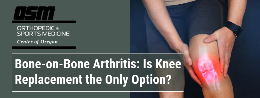
Bone-on-Bone Arthritis: Is Knee Replacement the Only Option?

Article featured on Orthopaedic Specialists

Many patients are told they have “bone-on-bone” arthritis, or end-stage osteoarthritis, on their x-ray and immediately worry that knee replacement surgery is their only option. The truth is, imaging alone should never determine whether you need surgery or not. The real indication for a knee replacement is pain that limits your daily activities. In addition, the suggestion for surgery should only come after you’ve completed a full course of conservative, non-operative treatment. If the pain is still unbearable, then imaging and discussions with your doctor can help you make the decision of whether or not to get knee replacement surgery.

Key Takeaway About a Bone-On-Bone Arthritis Diagnosis: An x-ray that shows severe arthritis does not automatically mean you need a knee replacement. Your symptoms and your response to conservative care matter far more than the image.

What Are Non-Operative Approaches to Bone-On-Bone Arthritis?

A comprehensive, non-operative approach can include treatments such as:

  • Physical therapy to improve strength and mechanics
  • Injection therapy such as corticosteroids or viscosupplementation to decrease inflammation
  • Bracing to unload painful areas
  • Activity modification to reduce stress on the joint.

Many patients experience significant improvement with these treatments, even with advanced arthritis.

What If I Still Experience Knee Pain After Non-Operative Approaches?

If, after diligently trying all these options, you are still struggling with pain, stiffness, or loss of function, then knee replacement becomes an excellent and appropriate solution. The surgery is designed to relieve pain and restore quality of life. But, it should be considered only when other methods have failed. It takes a long time to recover from knee replacement surgery and, like all surgeries, there are certain issues that can arise from it.

Allowing your body to heal itself naturally is often the best route. But, pain should never take away from your quality of life, especially with our advances in modern medicine.

Frequently Asked Questions About Bone-On-Bone Arthritis

  1. What can be done for bone-on-bone arthritis? Treatment options for osteoarthritis include non-operative methods like physical therapy, corticosteroid injections, braces or other assistance devices, and lifestyle changes. Surgical treatment can include a partial knee replacement or a full knee replacement surgery.
  2. What stage of arthritis is bone-on-bone arthritis? There is no medical diagnosis for ‘bone-on-bone”. The term simply describes late-stage, advanced, or severe osteoarthritis. Some medical experts may also refer to it as end-stage osteoarthritis.
  3. Is bone-on-bone arthritis serious? End-stage arthritis is only as serious as the pain level and frequency of flare-ups. If imaging tells you that you have ‘bone-on-bone arthritis’ but you haven’t experienced any unordinary levels of pain, or your arthritic flare-ups don’t bother you often, it might not be so serious as to consider surgery.
  4. Can you live with untreated bone-on-bone arthritis? Yes, of course! Osteoarthritis is the most common form of arthritis and affects a large portion of adults as they age. There are several ways to offset the symptoms and manage the condition that don’t require a doctor’s prescription or any medical intervention.

The Orthopedic & Sports Medicine Center of Oregon

The Orthopedic & Sports Medicine Center of Oregon (OSM) is an award-winning, board-certified orthopedic and sports medicine practice serving Lake Oswego, Portland, Scappoose, and surrounding Oregon communities. Our main clinic is located in Lake Oswego, with additional locations in Portland and Scappoose.

OSM provides comprehensive orthopedic care, sports medicine, spine care, joint replacement, foot and ankle surgery, hand and upper extremity care, and fracture treatment. Our physicians treat a wide range of conditions including sports injuries, arthritis, joint pain, spine conditions, ligament and tendon injuries, fractures, and degenerative musculoskeletal disorders using both surgical and nonsurgical approaches.

Our mission is to help patients return to pain-free movement, strength, and function through personalized treatment plans and advanced orthopedic techniques.


OSM Locations

Lake Oswego (Main Clinic)
17355 Lower Boones Ferry Rd, Suite 100A
Lake Oswego, OR 97035

Portland
5050 NE Hoyt St, Suite 668
Portland, OR 97213

Scappoose
51385 SW Old Portland Rd, Suite A
Scappoose, OR 97056


Phone: 503-224-8399
Hours: Mon–Thurs, 8:00am–4:30pm/ Friday 8:00am–1:00pm

If you are looking for experienced orthopedic surgeons, sports medicine specialists, spine doctors, or foot and ankle experts in Lake Oswego, Portland, or Scappoose, contact The Orthopedic & Sports Medicine Center of Oregon today.

What You Need to Know About Runner’s Knee

Article featured on WebMD

Runner‘s knee isn’t just for runners, and it’s not one particular injury. It’s a broad term that describes kneecap pain that can result from many different problems. You might hear a doctor call it patellofemoral pain syndrome.

Runner’s knee causes pain at the front of the knee, around the kneecap (also called the patella). It‘s common in people who run, jump, or play sports that put a lot of stress on the knees. Some people also call it jumper‘s knee.

But it can also happen to people who don’t play sports. Your kneecap can get out of line, causing it to move incorrectly in its groove on your thighbone (the femur). This can wear down the cartilage, a smooth, slippery layer that helps your bones glide easily against each other. That can cause pain.

You might feel a dull ache in your kneecap. It may hurt when you climb stairs, jump, squat, or after you sit for a long time. Some people also notice popping or crackling sounds when they stand up or go upstairs.

Here are some common triggers for runner’s knee:

Overuse. Bending your knees again and again or doing lots of high-stress exercises, such as lunges and plyometrics (training that uses the way your muscles lengthen and shorten to boost their power), can irritate tissues in and around your kneecap.A direct hit to the knee. Falling or bumping your knee can cause runner’s knee.Your bones aren’t aligned correctly (malalignment of the kneecap). If any of the bones from your hips to your ankles are out of their correct position, including the kneecap, that can put too much pressure on certain spots. Then, your kneecap can slip out of place and cause pain.

Problems with your feet that affect how you move. One such issue is called hypermobile feet, where the joints in and around your feet move more than they should. Other problems, such as flat feet or feet that roll inward when you walk, can also lead to knee pain.

Weak or unbalanced thigh muscles. The quadriceps, the big muscles in the front of your thigh, keep your kneecap in place when you bend or stretch the joint. If they‘re weak or tight, your kneecap may not stay in the right spot.Chondromalacia patella. This condition happens when the cartilage under your kneecap breaks down. When your kneecap doesn‘t glide smoothly, it can cause pain in the front of your knee.

Runner‘s knee can show up during or after physical activity and may get worse with certain movements. You might notice:

  • Pain at the front of the knee, especially around or under the kneecap
  • Tenderness along the inner edge of the kneecap
  • Swelling that appears after exercise
  • Stiffness or pain when walking up or down hills, or after sitting for a long time
  • Clicking, popping, or cracking sounds when you move your knee

To effectively treat your knee pain, your doctor needs to find out what caused it. They‘ll look at your knee, ask about your symptoms, and talk to you about your health history.

Your doctor may ask if the pain feels sharp or dull, and what makes it worse. They might press on your knee and watch how it moves when you walk, squat, or jump. This helps them check how well your knee moves, how strong it is, and where it hurts. They‘ll also look at how your knee lines up with the rest of your leg.

Sometimes, a physical exam is enough to tell if you have runner’s knee. If not, your doctor might order tests such as an X-ray or an MRI. In some cases, they may also use an ultrasound. They will try to rule out other problems, such as osteoarthritis (OA) or an unstable joint, that could be causing your knee pain.

For most people, runner’s knee gets better on its own with time, rest, and self-care. To help relieve your pain and speed recovery, you can:

Rest your knee. As much as possible, try to avoid things that make it hurt worse, such as running, squatting, lunging, or sitting and standing for long periods of time.

Ice your knee. This will help ease pain and swelling. Do it for 20-30 minutes every three to four hours for two to three days, or until the pain goes away.Wrap your knee. Use an elastic bandage, patellar straps, or sleeves to give it extra support.

Elevate your leg. Use a pillow to raise it when you sit or lie down.Take an over-the-counter anti-inflammatory medicine, such as ibuprofen or naproxen. These nonsteroidal anti-inflammatory drugs (NSAIDs) help with pain and swelling. But they can have side effects, such as a higher risk of bleeding and ulcers. Use as directed on the label, unless your doctor says otherwise.Do stretching and strengthening exercises. These are especially important for your quadriceps muscles. Your doctor can recommend a physical therapist to teach you what to do.

Try arch supports or orthotics for your shoes. They may help with the position of your feet. You can buy them at the store or get them custom-made.

If these techniques don’t help with your knee pain, ask your doctor if you need to see a specialist, such as an orthopedic surgeon. It‘s rare, but you could need surgery for a severe case of runner’s knee. An orthopedic surgeon can remove or replace damaged cartilage. In extreme cases, they can fix the position of your kneecap to spread stress more evenly across the joint.Knee braces for runner’s kneeA knee brace gives your knee support and keeps it stable to prevent extra movement or added stress. Some people use them for runner’s knee. But research on their effectiveness has had mixed results.

There are several different types of knee braces. Some that you might use for runner’s knee include:

  • Knee sleeves, which give you compression and a feeling of stability
  • Patellar tracking orthoses, which have straps to help keep your kneecap from moving out of line. Some also have hinges on the sides for extra stability.
  • Patellar straps, simple bands that use pressure to reduce strain on the tendon between your kneecap and shinbone

Knee braces are generally safe, but they can sometimes cause minor issues, such as skin irritation. A brace might not ease your symptoms or prevent injury. Talk to your doctor about what to expect and ask if other treatments might help.

People heal at their own pace. Your recovery time depends on your body and how serious your injury is.

While you get better, you need to take it easy on your knee. That doesn’t mean you have to stop all exercise. Just try activities that won’t strain your joints. For example, if you usually jog, try swimming instead.Whatever you do, don’t rush things. Resuming workouts too soon can cause more damage to your knee. Wait to return to your regular physical activities until:

  • You can fully bend and straighten your knee without pain.
  • You feel no pain in your knee when you walk, jog, sprint, or jump.
  • Your affected knee is as strong as your healthy knee.

You can’t always avoid runner’s knee, but you can lower your chances of getting it. Small changes in how you move or what you wear can help protect your knees:

  • Keep your thigh muscles strong and flexible with regular exercise.
  • Use shoe inserts if you have foot problems that may lead to runner’s knee.
  • Make sure your shoes have enough support.
  • Try not to run on hard surfaces, such as concrete.
  • Stay in shape and maintain a weight that’s healthy for you.
  • Warm up before you work out.
  • Don’t make sudden workout changes, such as adding squats or lunges. Add intense moves slowly.
  • Ask your doctor if you should see a physical therapist.
  • If your doctor or physical therapist suggests it, try a knee brace when you work out.
  • Wear quality running shoes.
  • Get a new pair of running shoes once yours lose their shape or the sole becomes worn or uneven.

The Orthopedic & Sports Medicine Center of Oregon is an award-winning, board-certified orthopedic group located in downtown Portland Oregon. We utilize both surgical and nonsurgical means to treat musculoskeletal trauma, spine diseases, foot and ankle conditions, sports injuries, degenerative diseases, infections, tumors and congenital disorders.

Our mission is to return our patients back to pain-free mobility and full strength as quickly and painlessly as possible using both surgical and non-surgical orthopedic procedures.

Our expert physicians provide leading-edge, comprehensive care in the diagnosis and treatment of orthopedic conditions, including total joint replacement and sports medicine. We apply the latest state-of-the-art techniques in order to return our patients to their active lifestyle.

If you’re looking for compassionate, expert orthopedic and podiatric surgeons in Portland Oregon, contact OSM today.

Phone:
503-224-8399

Address
17355 Lower Boones Ferry Rd Suite 100A
Lake Oswego, OR 97035

Hours
Monday–Friday
8:00am – 4:30pm

Getting Your Knees Ready for Summer Hiking

Article featured on AZ Ortho

Are you an avid hiker ready to hit the trails and conquer new heights? We get the appeal—hiking is an amazing way to connect with nature while staying active at the same time. However, you have to keep an eye on your joints, especially your knees, to ensure a smooth and pain-free hiking experience.

Here are some handy orthopedic tips specifically tailored for hikers to prevent joint strain and keep your knees trail-ready.

Choose the Right Footwear

Investing in the right footwear can make a significant difference in protecting your knees while hiking. Opt for hiking boots or shoes that are specifically designed for rugged terrain. Look for features like ankle support, cushioned insoles, and durable outsoles with good traction. Properly fitting footwear also helps prevent blisters, hot spots, and discomfort that can indirectly affect your knee health.

Additional Tip: Consider Orthotic Inserts

If you have specific foot issues or pronation concerns, consider using orthotic inserts in your hiking shoes. Orthotics can provide additional support and alignment for your feet, which can help reduce stress on your knees.

Warm-Up and Stretch

Before starting your hike, spend at least 10–15 minutes warming up your muscles and performing dynamic stretches. No one should go into any type of exercise with cold muscles!

Good dynamic stretches should involve active movements that mimic the actions you’ll perform during the hike, such as leg swings, high knees, and lunges. These exercises increase blood flow to your muscles, improve flexibility, and prepare your body for the physical demands of hiking.

Additional Tip: Include Foam Rolling

Incorporate foam rolling into your pre-hike routine to release tension in your muscles and fascia. Focus on rolling out your quadriceps, hamstrings, calves, and IT band. Foam rolling can help improve your range of motion, reduce muscle tightness, and enhance overall mobility, which can contribute to better knee health during your hike.

Maintain Proper Posture

Proper posture lets you prevent knee strain and maintain good overall body alignment during hiking. Keep your head up, shoulders relaxed, and gaze forward to avoid rounding your shoulders or slouching. Engage your core muscles to support your spine and pelvis, which reduces strain on your lower back and knees. Periodically check your posture throughout your hike and make adjustments as needed.

Additional Tip: Use Backpack Ergonomics

If you’re carrying a backpack during your hike, make sure the straps are adjusted properly to distribute weight evenly. Use a backpack that has padded shoulder straps, a waist belt, and a chest strap to stabilize the load and prevent strain on your back and shoulders. Pack essential items strategically to minimize unnecessary weight and optimize balance while hiking.

Gradually Increase Intensity

Whether you’re a beginner or an experienced hiker, it’s best to progress gradually when increasing the intensity of your hikes. Start with shorter distances and easier trails, gradually building up to longer hikes with more elevation gain. Listen to your body’s cues and take breaks as needed to rest and refuel. Pushing yourself too hard too soon can lead to overuse injuries and trouble for your knees.

Additional Tip: Incorporate Cross-Training

Incorporate cross-training activities like cycling, swimming, or yoga into your fitness routine to gain better strength, flexibility, and endurance. Cross-training can help prevent muscle imbalances, improve joint stability, and reduce the risk of overuse injuries.

Watch Your Foot Placement

Pay attention to where you step while hiking to avoid uneven terrain and obstacles that could lead to slips or falls. Take deliberate steps and use caution when navigating rocky or slippery surfaces. Choose stable footing whenever possible and use trekking poles for added balance and support, especially during descents or challenging terrain.

Additional Tip: Use Proper Descending Techniques

When descending steep slopes or rocky terrain, use proper techniques to protect your knees. Descend slowly and maintain a slightly bent knee position to absorb shock and reduce the impact on your joints. Avoid locking your knees or landing with excessive force, as this can strain your knee ligaments and cartilage.

Strengthen Your Leg Muscles

Strong leg muscles provide the support and stability your knees need during hikes. Incorporate strength training exercises into your fitness routine to target your quadriceps, hamstrings, calves, and glutes. Squats, lunges, calf raises, and leg presses are excellent exercises to build strength and improve knee stability. Consistent strength training can help prevent joint strain and enhance your hiking performance.

Additional Tip: Focus on Functional Movements

In addition to traditional strength training exercises like squats, lunges, and leg presses, prioritize functional movements that mimic hiking actions. Include exercises such as step-ups, single-leg squats, and lateral lunges to improve balance, stability, and agility. Functional training prepares your muscles for the specific demands of hiking, reducing the risk of injuries.

Listen to Your Body

Lastly, listen to your body and pay attention to any signs of discomfort or pain in your knees while hiking. If you experience persistent pain, swelling, or instability, seek medical attention and avoid pushing through the pain. Rest, ice, compression, and elevation (RICE) can help alleviate minor knee injuries, but consulting with an orthopedic specialist is needed for proper diagnosis and treatment.

Additional Tip: Implement Recovery Strategies

After your hike, prioritize recovery strategies to promote muscle recovery and reduce inflammation in your knees. Perform gentle stretching exercises, use foam rollers or massage tools to alleviate muscle tension, and apply ice packs to reduce swelling. To get fully recovered and help your joints, get a good amount of rest, hydration, and nutrition as well.


The Orthopedic & Sports Medicine Center of Oregon is an award-winning, board-certified orthopedic group located in downtown Portland Oregon. We utilize both surgical and nonsurgical means to treat musculoskeletal trauma, spine diseases, foot and ankle conditions, sports injuries, degenerative diseases, infections, tumors and congenital disorders.

Our mission is to return our patients back to pain-free mobility and full strength as quickly and painlessly as possible using both surgical and non-surgical orthopedic procedures.

Our expert physicians provide leading-edge, comprehensive care in the diagnosis and treatment of orthopedic conditions, including total joint replacement and sports medicine. We apply the latest state-of-the-art techniques in order to return our patients to their active lifestyle.

If you’re looking for compassionate, expert orthopedic and podiatric surgeons in Portland Oregon, contact OSM today.

Phone:
503-224-8399

Address
17355 Lower Boones Ferry Rd Suite 100A
Lake Oswego, OR 97035

Hours
Monday–Friday
8:00am – 4:30pm

Common Swimming Overuse Injuries

Article featured on Physiopedia

Swimming is a great sport enjoyed by all age groups at all levels of ability. It is a unique sport, in that it combines upper and lower extremity strength exercises with cardiovascular training in a non-weight bearing environment. Swimmers are unfortunately prone to injuries. Most swimming injuries are classed as overuse injuries and relate to faulty biomechanics. The most common swimming injury regions are the shoulder, neck, lower back and knee.

Swimming overuse injuries usually arise from one or a combination of the below:

  • Poor stroke mechanics
  • Poor breathing technique
  • Poor flexibility or range of motion of neck or lower back
  • Hyper flexibility of joints with insufficient muscular stabilisation
  • Decreased rotator cuff or scapular muscle strength
  • Insufficient core strength/stability
  • Decreased hip muscle strength
  • Overtraining
  • Insufficient rest periods

Stretching is important for swimming because of the great range of muscles used when you’re in the water.  Unprepared muscles don’t perform as well as muscles which have been warmed up ahead of exercising. A good stretching regime including at a minimum:

  • posterior shoulder stretch
  • pectoral stretch
  • long head biceps stretch
  • upper thoracic spine mobility stretch
  • latissimus dorsi

Note – The above list gives a guide to what to look for in each of the specific conditions listed below. This page does not give full physiotherapy procedures, instead providing great links to the sites that will have relevant information.

Swimmers Shoulder

Shoulder pain is the most frequent orthopaedic injury in swimmers, with a reported prevalence between 40% and 91% in elite swimmers. Swimmer’s shoulder is a condition with a gradual onset due to repetitive activity and can be classified as microtrauma. Swimmer’s shoulder usually presents as subacromial impingement involving the rotator cuff tendon, bicipital tendon, or subacromial bursa.

Primary subacromial impingement involves compression of these structures between the acromion and greater tuberosity (due to usually a tight posterior capsule causing the humeral head to migrate anteriorly) or abnormal acromial morphology. However, primary impingement syndrome is less common in competitive swimmers than secondary impingement.

Secondary impingement occurs through a series of impairments, usually in a swimmer with increased anterior glenohumeral laxity (shoulder ROM in swimmers often exhibit excessive external rotation and limited internal rotation). This shift in ROM towards increased external rotation is an adjustment to the demands on the glenohumeral joint allowing anterior laxity and greater demand on the rotator cuff and the long head of the biceps to reduce humeral head elevation and anterior translation.

Chronic swimmer’s shoulder can result in pathology of the rotator cuff, glenoid labrum, and long head of the biceps.

Swimmers Back

The spine is also a recognized site predisposed to injury in the elite swimmer, mainly associated with butterfly swimmers ( 33.3%-58%) and breaststroke swimmers (22.2%- 47%), figures varying from differing studies.

As swimming athletes are positioned horizontally in the water, gravity and buoyancy forces are traveling through the body with the potential to cause changes at the lumbar spine. Due to the undulating motion (lumbar extension) utilised in butterfly and breaststroke, there is potential for developing pain secondary to stress on the joints.

Causes include:

  • Hyperextension of the lumbar spine during freestyle and butterfly
  • poor kick technique
  • stress fractures are a potential cause in young swimmers
  • disc degeneration and facet joint degeneration in the older swimmer. A 2007 study hypothisising that “Excessive competitive swimming activities accelerate lumbar disk degeneration” this activity may exaggerate lumbar intervertebral disk degeneration, especially in the L5-S1 intervertebral segment.
  • improper timing of this butterfly stroke increases strain on low back the possibility of neck, shoulder, or back pain.
  • poor flexibility of the spine and low back
  • insufficient core strength to maintain straight alignment of body in water
  • aggressive weight training using poor techniques
  • overuse of devices e.g. paddles on hands, fins, kick boards- exposing the open kinetic chain of swimming to different loads and exaggerating lumbar lordosis
  • tightness in the hip flexors or inadequate body roll during swimming may lead to compensation at the lumbar spine.

Swimmers Knee

Knee pain figures range from 34% to 86% for a single knee episode, being highest in breaststrokers.

Knee pain is caused mainly during breaststroke swimming. Repetitive stress is placed on the medial knee produces pain during the whip-like motion. The breaststroke kick is a high valgus load produced during sudden flexion-extension, adduction and external rotation of the knee against the hydrodynamic environment, resulting in stress to the medial compartment. Hence a strain occurs to the medial collateral ligament and compression on the lateral knee (possibly causing sprain on the MCL; irritation of the medial plica; bursal irritation at the muscular insertions of the adductor and hamstring muscles). Strain injuries may also present in the adductor muscles (adductor magnus and brevis, especially). Other knee injuries include patellofemoral pain, and medial synovitis. Treatment will focus on elimination of inflammation. Rehabilitation should focus on stabilisation exercises for hypermobile joints, postural correction, strengthening and flexibility.

A 2008 study found the 200 – 400 m breaststroke events increase the risk for knee overuse injuries more than other strokes or distances. Additionally training for more than four times a week increases the risk twofold for knee and fourfold for shoulder overuse injuries.

A 2004 study looking at competitive swimmers interestingly reports that breaststroke swimmers are at significant risk of: groin injury; groin injury is positively correlated with increased magnitude of breaststroke training; and groin injury may prevent participation in practices and competitions. So take the time to assess then groin and complete length of hip adductors, not just distally.

Swimmers Neck

Neck pain is mainly seen in the older athlete It may be due to facet joint arthritic change, and disc degeneration +/- nerve root irritation.  Muscle innervation and sensation to the shoulder region is predominantly derived from the C5/C6 nerve roots.  If these nerve roots are irritated due to degenerative change in the neck, shoulder complaints can arise.  Arthritic change may limit neck rotation making correct breathing patterns difficult.  Swimmers who unilaterally breath are more prone to neck pain.  Looking forward rather than directly downward and extending the head too high when taking a breath increases the load on the neck and create pain.

Conclusion

Swimming can be a great sport for recreational swimmers up to elite swimmers. As a physiotherapist help swimmers overcome their injuries and liaise with trainers, coaches and physicians for the best results.


The Orthopedic & Sports Medicine Center of Oregon is an award-winning, board-certified orthopedic group located in downtown Portland Oregon. We utilize both surgical and nonsurgical means to treat musculoskeletal trauma, spine diseases, foot and ankle conditions, sports injuries, degenerative diseases, infections, tumors and congenital disorders.

Our mission is to return our patients back to pain-free mobility and full strength as quickly and painlessly as possible using both surgical and non-surgical orthopedic procedures.

Our expert physicians provide leading-edge, comprehensive care in the diagnosis and treatment of orthopedic conditions, including total joint replacement and sports medicine. We apply the latest state-of-the-art techniques in order to return our patients to their active lifestyle.

If you’re looking for compassionate, expert orthopedic and podiatric surgeons in Portland Oregon, contact OSM today.

Phone:

503-224-8399

Address
17355 Lower Boones Ferry Rd Suite 100A
Lake Oswego, OR 97035

Hours
Monday–Friday
8:00am – 4:30pm

What Are ACL, MCL, and PCL Injuries?

Article featured on Seattle Children’s Hospital

What are ACL, MCL and PCL injuries?

ACL, MCL and PCL injuries are damage to ligaments that keep the knee from wobbling or giving out when you move. These ligaments are the anterior cruciate ligament (ACL), medial collateral ligament (MCL) and posterior cruciate ligament (PCL). When the knee is forced into an unusual position, 1 or more of the ligaments can tear, either part way or all the way.

What are the symptoms of ACL, MCL and PCL injuries?

If the knee of a child, teen or young adult suddenly swells after an accident or injury, they may have a torn ACL, MCL or PCL or another serious knee problem. Other symptoms of knee ligament injuries include:

  • A popping sound at the time of the injury
  • Pain in the knee
  • Knee is wobbly or unstable

How are ACL, MCL and PCL injuries diagnosed?

To help prevent further injuries to the bones and joints, it is important to get a thorough evaluation and proper treatment.

At our clinic, the doctor will:

  • Ask what happened before the knee began to swell and hurt.
  • Examine the knee. To doctors trained in sports medicine, knees with ACL, MCL or PCL tears often feel loose.

We cannot be sure that a knee ligament is torn until the swelling in the knee has gone down. This usually takes 7 to 10 days. At that time, we may take:

  • X-rays of the knee to get more information on what is wrong
  • An MRI if it is not clear that a ligament is torn or if we think there may be other injuries

How are ACL, MCL and PCL injuries treated?

Young people with a torn ACL need surgery to repair or reconstruct the ligament and then physical therapy (PT) to help them get back to their usual activities. If they are still growing, it’s important to receive care from a pediatric orthopedic surgeon who knows how to protect their .

For MCL and PCL injuries, we use nonsurgical methods like PT, including sports PT, whenever possible. Those who have an MCL injury may need to wear a hinged knee brace along with doing PT.

Surgery for ACL, MCL and PCL injuries

Surgery to repair a torn ACL, called transepiphyseal anterior cruciate ligament reconstruction. The red lines are the new ligament. The black dots show where the surgeon secures the new ligament to the thighbone above the knee and the shinbone below the knee.

Surgery is the usual treatment after an ACL injury to make the knee stable again.

During ACL surgery, the surgeon replaces the torn ligament with new tissue that will turn into ligament over time. This tissue (called a graft) is taken from another body site and transferred to the new area. Graft tissue can be taken from the patient’s own body or from a donor (cadaver). The surgeon will talk with you in advance about the pros and cons of different types of grafts.

We drill small holes in the shinbone and the thighbone. Then, we pass the new ligament through the drill holes and secure it to the bones.

This surgery requires only small incisions. The surgeon inserts a small tool called an arthroscope into the knee. They use pictures displayed on a large monitor to guide their actions.

A young person who has both an MCL injury and an ACL injury probably will need surgery to reconstruct the ACL.

Surgery is not usually used to treat PCL injuries that happen by themselves. But surgery may be needed if:

  • A PCL injury happens along with an injury to the ACL or another part of the knee.
  • An injury moves the piece of bone the PCL is attached to far from its usual place on the shinbone. Surgeons can put the bone back and attach it again.

Physical therapy for ACL, MCL and PCL injuries

PT is an important part of getting better after surgery for any knee ligament injury. It will help strengthen the muscles around the knee and make the joint stable.

If an MCL or PCL injury is mild, a child, teen or young adult may be able to recover with PT alone. Our staff is expert at devising and teaching exercise programs to help patients:

  • Recover range of motion
  • Rebuild strength and stability
  • Restore balance
  • Regain confidence in using their knee

Most patients will have PT sessions with us a couple of times a week until they meet their goals for returning to their sport or other usual activities. Afterward, our therapists may also recommend exercises to keep doing at home.

To prevent new or repeat injuries, we have a strong focus on return-to-sport testing. Our PTs know how to check when a patient is ready to be more active or go back to their sport. We have done a great deal of research to understand how to make these decisions so each patient gets the best possible results.

One of the most important ways to prevent repeat injuries is to modify activities that might hurt the knee as it heals. These activities include high-level athletics that require:

  • Jumping
  • Pivoting
  • Contact with other players
  • Playing on an uneven surface

The Orthopedic & Sports Medicine Center of Oregon is an award-winning, board-certified orthopedic group located in downtown Portland Oregon. We utilize both surgical and nonsurgical means to treat musculoskeletal trauma, spine diseases, foot and ankle conditions, sports injuries, degenerative diseases, infections, tumors and congenital disorders.

Our mission is to return our patients back to pain-free mobility and full strength as quickly and painlessly as possible using both surgical and non-surgical orthopedic procedures.

Our expert physicians provide leading-edge, comprehensive care in the diagnosis and treatment of orthopedic conditions, including total joint replacement and sports medicine. We apply the latest state-of-the-art techniques in order to return our patients to their active lifestyle.

If you’re looking for compassionate, expert orthopedic and podiatric surgeons in Portland Oregon, contact OSM today.

Phone:

503-224-8399

Address
17355 Lower Boones Ferry Rd Suite 100A
Lake Oswego, OR 97035

Hours
Monday–Friday
8:00am – 4:30pm