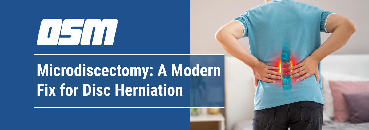
Microdiscectomy: A Modern Fix for Disc Herniation

What is microdiscectomy?

Microdiscectomy is a surgical procedure for the relief of pain and other symptoms that occur when a herniated disc in the spine presses on an adjacent nerve root. During the operation, the surgeon frees the nerve by removing small fragments of disc, bone and ligament.

Microdiscectomy is sometimes described as a minimally invasive spine surgery, because it requires only a small incision and the use of a microscope or surgical glasses, called loupes, to magnify the site where the injury has occurred. The surgeon also uses smaller tools and instruments to work in the restricted space of the spine. Microdiscectomy is a type of spinal decompression surgery, and the term microdecompression – which describes the surgical removal of any bone or ligament that is compressing a nerve – may also be used to describe the procedure. However, the term “microdiscectomy” is more specific to the removal of the fragment of herniated disc tissue that is causing the patient’s symptoms.

Because most patients will recover from a herniated disc without surgery, microdiscectomy is recommended only after conservative treatment, including physical therapy, cortisone shots, and other medication have been tried for a period of at least 6 to 12 weeks, without bringing relief. In some cases, motor weakness may be a reason to have this surgery sooner. Patients with cauda equina syndrome require immediate surgical intervention. This is a condition in which pressure on the nerves in the lower portion of the spine affects bladder and bowel function, but it affects less than 1% of people.

How does a surgeon perform a microdiscectomy?

Microdiscectomy surgery may be approached using one of three minimally invasive techniques to gain access to the herniated disc and nerve: a midline, tubular or endoscopic microdiscectomy.

Historically, a traditional discectomy required a large incision and involved removal of the entire disc. Today, with the availability of advanced techniques and equipment, almost all spine surgeons perform microdiscectomies. The procedure usually results in rapid, and sometimes immediate, pain relief.

Microdiscectomy techniques

  • A midline microdiscectomy, in which the surgeon makes a one to two-inch vertical incision in the back, lifts the surrounding muscles off the vertebrae, and uses instruments to hold apart the layers of tissue during the procedure.
  • A tubular microdiscectomy, in which the surgeon inserts a series of small tubes or dilators through a small incision to create a corridor through the muscle in which to operate, thereby causing less disruption to this tissue.
  • Endoscopic microdiscectomy (sometimes also known as microendoscopic discectomy), in which an even smaller incision is made and the surgeon employs a miniaturized camera and instruments; this approach also involves less disruption to surrounding tissues.

What are the benefits of a microdiscectomy?

Regardless of which particular microdiscectomy technique selected, the surgical goal is the same: to remove the disc fragment and any bone or ligament that may be compressing the nerve root. To do so, the surgeon creates a small window in the vertebra (a procedure also called a laminotomy) and pulls away the ligamentum flavum, the underlying tissue, to reveal the dura (the covering of the spine) and the nerve root. The herniated part of the disc is then removed to complete the nerve decompression.

Imaging techniques, including X-rays, are used before, and sometimes during, the surgery to ensure localization of the correct operative site.

While herniated discs may occur in almost any level of the spine, lumbar microdiscectomy – surgery in the lower portion of the back – is the most common site for this procedure.

How long does a microdiscectomy take?

On average, microdiscectomy surgery takes between 30 to 60 minutes to complete. However, because patients are given general anesthesia and must spend time afterward in the recovery room for a period of monitoring, the total time usually extends to about two hours.

In almost all cases, people undergoing microdiscectomy may return home the same day. Upon discharge from the hospital, patients are typically given a small amount of pain medication – including acetaminophen or small dose of opioid medication – and a muscle relaxant with instructions regarding postsurgical care and how to taper these medications appropriately. Special emphasis is given to symptoms that signal the need for immediate medical attention, including muscle weakness, change in bladder or bowel function, and extreme and unexpected back or leg pain.

What complications can occur with microdiscectomy?

During surgery, multiple safety measures are followed to control bleeding, prevent infection and avoid injury to any surrounding tissues. Rarely, a tear in the dura (the tissue surrounding the spinal nerves) may occur. The surgeon repairs this with a suture or a patch made of collagen.

What is the expected microdiscectomy recovery time?

Following a two-week period of rest to allow the soft tissues to heal, many patients feel well enough to return to work. However, microdiscectomy recovery typically requires a six-week period of modified activity and includes a course of physical therapy starting at week two or three.

Physical therapists focus on helping patients strengthen core muscles and loosen stiff joints, as well as creating a home exercise program to help protect the spine. All patients are cautioned to ease back into their previous routines gradually, especially those whose work requires physical labor. Because sitting in a chair and leaning forward places pressure on the spine, people with more sedentary jobs also need to be attentive to their posture and take a short walk or do some gentle exercise every hour or so.

Patients who plan to resume driving after surgery must wait until they are pain-free, finished with any medication that causes drowsiness, and able to both shift their foot between pedals and turn in their seats easily and without pain. To test individual comfort, a practice drive in a parking lot or other safe space, in the company of another driver is recommended before returning to regular roadways.

Is a microdiscectomy painful?

Following surgery, most patients do well with a small amount of non-opioid pain medication and a drug that relaxes the muscles. While there is some discomfort associated with the surgical incision, many patients experience rapid relief of the pain caused by the herniated disc.

In patients who have experienced nerve compression and associated symptoms over an extended period, pain relief may take longer. When leg pain has been the primary symptom, the pain typically retreats up the leg over time. In addition, patients may experience muscle cramping or mild numbness or tingling after surgery which typically resolves with time.

Who is eligible for microdiscectomy?

Most patients with herniated discs that do not respond to a trial of medication and physical therapy over time, are eligible for a microdiscectomy. While the condition is usually seen in individuals aged 30 to 50, it does occur outside this age range.

Herniated discs are rare in children and young adults, who are more frequently able to recover without surgical intervention. Microdiscectomy may be appropriate for adults in their 80s or 90s, though they should be cautioned that there is an increased chance of medical or surgical complications in this population.

How successful is microdiscectomy?

Overall, microdiscectomy success rates are excellent, with many patients expressing a high degree of satisfaction with the outcome. Careful presurgical screening and evaluation, as well as patient commitment to maintaining good spine health following microdiscectomy, contribute to the success of the surgery.

It should be noted that some people with herniated discs are also found to have other issues in the spine that are causing nerve-related pain and disability. In such cases, additional procedures such as a laminectomy – removal of the lamina (bony roof over the spinal canal) in one or more vertebrae, may be required if it is felt that these issues are contributing to their disability.

Can microdiscectomy be used on a disc that herniates a second time?

In cases of disc reherniation, microdiscectomy can be performed for a second time on the same disc, often with good results. However, if herniation occurs in the same disc a third time, a different type of treatment will be recommended.


The Orthopedic & Sports Medicine Center of Oregon is an award-winning, board-certified orthopedic group located in downtown Portland Oregon. We utilize both surgical and nonsurgical means to treat musculoskeletal trauma, spine diseases, foot and ankle conditions, sports injuries, degenerative diseases, infections, tumors and congenital disorders.

Our mission is to return our patients back to pain-free mobility and full strength as quickly and painlessly as possible using both surgical and non-surgical orthopedic procedures.

Our expert physicians provide leading-edge, comprehensive care in the diagnosis and treatment of orthopedic conditions, including total joint replacement and sports medicine. We apply the latest state-of-the-art techniques in order to return our patients to their active lifestyle.

If you’re looking for compassionate, expert orthopedic and podiatric surgeons in Portland Oregon, contact OSM today.

Phone:

503-224-8399

Address
1515 NW 18th Ave, 3rd Floor
Portland, OR 97209

Hours
Monday–Friday
8:00am – 4:30pm

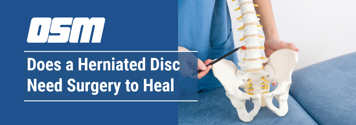
Does a Herniated Disc Need Surgery to Heal?


The Orthopedic & Sports Medicine Center of Oregon is an award-winning, board-certified orthopedic group located in downtown Portland Oregon. We utilize both surgical and nonsurgical means to treat musculoskeletal trauma, spine diseases, foot and ankle conditions, sports injuries, degenerative diseases, infections, tumors and congenital disorders.

Our mission is to return our patients back to pain-free mobility and full strength as quickly and painlessly as possible using both surgical and non-surgical orthopedic procedures.

Our expert physicians provide leading-edge, comprehensive care in the diagnosis and treatment of orthopedic conditions, including total joint replacement and sports medicine. We apply the latest state-of-the-art techniques in order to return our patients to their active lifestyle.

If you’re looking for compassionate, expert orthopedic and podiatric surgeons in Portland Oregon, contact OSM today.

Phone:

503-224-8399

Address
1515 NW 18th Ave, 3rd Floor
Portland, OR 97209

Hours
Monday–Friday
8:00am – 4:30pm


10 Common Back Pain Myths


The Orthopedic & Sports Medicine Center of Oregon is an award-winning, board-certified orthopedic group located in downtown Portland Oregon. We utilize both surgical and nonsurgical means to treat musculoskeletal trauma, spine diseases, foot and ankle conditions, sports injuries, degenerative diseases, infections, tumors and congenital disorders.

Our mission is to return our patients back to pain-free mobility and full strength as quickly and painlessly as possible using both surgical and non-surgical orthopedic procedures.

Our expert physicians provide leading-edge, comprehensive care in the diagnosis and treatment of orthopedic conditions, including total joint replacement and sports medicine. We apply the latest state-of-the-art techniques in order to return our patients to their active lifestyle.

If you’re looking for compassionate, expert orthopedic and podiatric surgeons in Portland Oregon, contact OSM today.

Phone:

503-224-8399

Address
1515 NW 18th Ave, 3rd Floor
Portland, OR 97209

Hours
Monday–Friday
8:00am – 4:30pm


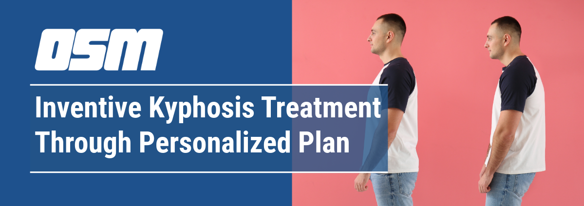
Inventive Kyphosis Treatment Through Personalized Plan

Article featured on Rothman Orthopedics

Kyphosis, commonly referred to as a forward curvature of the spine, is a condition that can cause significant discomfort and affect the quality of life. It can result from various factors, including poor posture, spinal abnormalities, or conditions such as osteoporosis. Fortunately, advancements in medical science and personalized treatment plans are revolutionizing the way we address this condition. In this blog post, we will delve into the journey of kyphosis correction and the pioneering personalized treatment plans that are transforming patient outcomes.

Understanding Kyphosis: More Than Just a Hunch

Kyphosis presents as an exaggerated forward rounding of the upper back, often leading to a noticeable hunch. While mild kyphosis may not cause major issues, severe cases can result in pain, breathing difficulties, and limited physical function. This condition can affect individuals of all ages, though it is particularly prevalent among older adults and adolescents experiencing growth spurts.

The Importance of Early Diagnosis

Early diagnosis is crucial in managing kyphosis effectively. Comprehensive evaluations involving physical examinations, patient history, and imaging studies (like X-rays or MRIs) help in determining the severity and underlying causes of the curvature. Recognizing the condition early allows for timely intervention, which can prevent further progression and complications.

Personalized Treatment Plans: A Game Changer

No two patients are the same, and neither are their kyphosis conditions. Personalized treatment plans consider the individual’s specific condition, overall health, and lifestyle. This tailored approach ensures that each patient receives the most effective and appropriate care. Here’s how personalized treatment plans are transforming kyphosis correction:

  • Holistic Assessment: A thorough assessment of the patient’s medical history, lifestyle, and specific kyphosis characteristics forms the foundation of a personalized treatment plan. This holistic approach allows for a comprehensive understanding of the patient’s needs. Factors such as the severity of the curvature, the patient’s age, activity level, and overall health are all considered to create a plan that is uniquely suited to the individual.
  • Non-Surgical Interventions: For many patients, non-surgical options are the first line of defense. These may include physical therapy to strengthen the back muscles, improve posture, and increase flexibility. Bracing can also be effective, especially in younger patients whose bones are still growing. Customized exercise programs and ergonomic adjustments can also play a significant role in managing symptoms and improving quality of life.
  • Minimally Invasive Procedures: Advances in medical technology have led to the development of minimally invasive procedures that can correct kyphosis with reduced recovery times and fewer complications. Techniques such as vertebroplasty or kyphoplasty involve stabilizing the spine with small incisions, offering a quicker return to normal activities. These procedures are particularly beneficial for older adults or those with osteoporosis, where traditional surgery may carry higher risks.
  • Surgical Solutions: In severe cases where non-surgical methods are insufficient, surgery may be required. Modern surgical techniques are highly precise, aiming to correct the curvature while minimizing risks. Personalized surgical plans ensure that each patient’s unique anatomy and needs are considered, leading to better outcomes. Advanced surgical techniques, including spinal fusion and osteotomy, are tailored to correct the specific type and location of the curvature.
  • Post-Treatment Rehabilitation: Recovery doesn’t end with treatment. Personalized rehabilitation programs, including physical therapy and lifestyle modifications, are essential for maintaining the correction and preventing recurrence. Continuous monitoring and follow-ups ensure that the patient’s progress is on track. Rehabilitation may also involve pain management strategies and patient education to promote long-term spinal health.

The Orthopedic & Sports Medicine Center of Oregon is an award-winning, board-certified orthopedic group located in downtown Portland Oregon. We utilize both surgical and nonsurgical means to treat musculoskeletal trauma, spine diseases, foot and ankle conditions, sports injuries, degenerative diseases, infections, tumors and congenital disorders.

Our mission is to return our patients back to pain-free mobility and full strength as quickly and painlessly as possible using both surgical and non-surgical orthopedic procedures.

Our expert physicians provide leading-edge, comprehensive care in the diagnosis and treatment of orthopedic conditions, including total joint replacement and sports medicine. We apply the latest state-of-the-art techniques in order to return our patients to their active lifestyle.

If you’re looking for compassionate, expert orthopedic and podiatric surgeons in Portland Oregon, contact OSM today.

Phone:

503-224-8399

Address
1515 NW 18th Ave, 3rd Floor
Portland, OR 97209

Hours
Monday–Friday
8:00am – 4:30pm

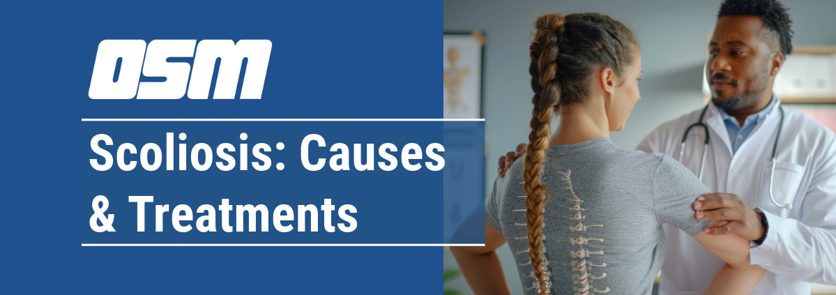
Scoliosis: Causes and Treatments

Article featured on Johns Hopkins Medicine

What is scoliosis?

The spine is made up of a stack of rectangular-shaped building blocks called vertebrae. When viewed from behind, the spine normally appears straight. However, a spine affected by scoliosis is curved — often appearing like an S or C — with a rotation of the vertebrae. This curvature gives the appearance that the person is leaning to one side.

Scoliosis is determined when the curvature of the spine measures 10 degrees or greater on an X-ray. Spinal curvature from scoliosis may occur on the right or left side of the spine, or on both sides in different sections. Both the thoracic (mid) and lumbar (lower) spine may be affected by scoliosis. Scoliosis is a type of spinal deformity.

In more than 80 percent of cases, the cause of scoliosis is unknown — a condition called idiopathic scoliosis. In other cases, scoliosis may develop as a result of degeneration of the spinal discs, as seen with arthritis, osteoporosis or as a hereditary condition that tends to run in families.

What are the different types of scoliosis?

Congenital Scoliosis

In congenital scoliosis, spinal curvature develops because of misshapen vertebrae. The diagnosis of congenital scoliosis may be made in early infancy if outward signs are present, but many cases are diagnosed later in childhood.

As a child grows, scoliosis may worsen, and asymmetries in the body may develop. Typically, congenital scoliosis is treated with a “watch and wait” approach. Surgery is considered only if a curve is clearly getting worse and the child is facing ongoing deformity and risk of future pain.

Idiopathic Scoliosis

Doctors, nurses and scientists have been studying the natural history and genetics of scoliosis for decades, but to this day, the cause of idiopathic scoliosis is still unknown. But we do know that the most common time for idiopathic scoliosis to develop is at the onset of adolescence, or around the age of 10. We also know that growth can make it worse, and we should be most concerned about scoliosis in a child that has significant growth remaining.

When diagnosed in children 2 or younger, this type of scoliosis is called infantile idiopathic scoliosis.

Neuromuscular Scoliosis

A child with an underlying neuromuscular condition is at higher risk for developing scoliosis. A straight spine requires normal muscle balance and strength in the torso. In conditions such as cerebral palsy, spina bifida and muscular dystrophy, the muscles are often weak and unbalanced, leading to the development of a spinal curvature.

A child with neuromuscular scoliosis is given the option of wearing a scoliosis brace that may slow or prevent the worsening of the condition. Surgical intervention is offered when the curve has reached the tipping point of 50 degrees. Over time, these curves will continue to worsen, leading to progressive imbalance of the torso. Beyond 80 degrees, breathing challenges develop as space for the lungs decreases.

What are the symptoms of scoliosis?

The following are the most common symptoms of scoliosis. However, each individual may experience symptoms differently. Symptoms may include:

  • Difference in shoulder height
  • The head isn’t centered with the rest of the body
  • Difference in hip height or position
  • Difference in shoulder blade height or position
  • When standing straight, difference in the way the arms hang beside the body
  • When bending forward, the sides of the back appear different in height
  • Prominence or asymmetry in the ribs seen from the front or back

The symptoms of scoliosis may resemble other spinal conditions or deformities, or may be a result of an injury or infection. Always consult your doctor for a diagnosis.

Symptoms that are not commonly associated with idiopathic scoliosis are back pain, leg pain, and changes in bowel and bladder habits. If a person is experiencing these types of symptoms, he or she requires immediate further medical evaluation by a doctor to determine the cause of the symptoms.

How is scoliosis diagnosed?

Early detection of scoliosis is most important for successful treatment. In addition to a complete medical history and physical examination, an X-ray is the primary diagnostic tool for scoliosis. In establishing a diagnosis of scoliosis, the doctor measures the degree of spinal curvature on the X-ray.

The following additional diagnostic procedures may be performed for nonidiopathic curvatures, atypical curve patterns or congenital scoliosis:

  • MRI. This diagnostic procedure uses a combination of large magnets and a computer to produce detailed images of organs and structures within the body.
  • CT scan. This diagnostic imaging procedure uses a combination of X-rays and computer technology to produce horizontal, or axial, images (often called slices) of the body. A CT scan shows detailed images of any part of the body, including the bones, muscles, fat and organs. CT scans are more detailed than general X-rays.

How is scoliosis treated?

The goal of treatment is to stop the progression of the curve and prevent deformity. Observation and repeated examinations — also referred to as the “watch and wait” approach — may be necessary to determine if the spine is continuing to curve. These are used when a person has a curve that is less than 20 degrees and who is still growing.

For actively growing children with scoliosis curves between 20 and 50 degrees, bracing is recommended. An external torso brace, or TLSO, is worn for a prescribed number of hours. The brace applies corrective pressure to the growing spine, preventing further worsening of the scoliosis. Surgery is a recommended treatment option for a child with severe scoliosis or a curve that has worsened to more than 50 degrees.


The Orthopedic & Sports Medicine Center of Oregon is an award-winning, board-certified orthopedic group located in downtown Portland Oregon. We utilize both surgical and nonsurgical means to treat musculoskeletal trauma, spine diseases, foot and ankle conditions, sports injuries, degenerative diseases, infections, tumors and congenital disorders.

Our mission is to return our patients back to pain-free mobility and full strength as quickly and painlessly as possible using both surgical and non-surgical orthopedic procedures.

Our expert physicians provide leading-edge, comprehensive care in the diagnosis and treatment of orthopedic conditions, including total joint replacement and sports medicine. We apply the latest state-of-the-art techniques in order to return our patients to their active lifestyle.

If you’re looking for compassionate, expert orthopedic and podiatric surgeons in Portland Oregon, contact OSM today.

Phone:

503-224-8399

Address
1515 NW 18th Ave, 3rd Floor
Portland, OR 97209

Hours
Monday–Friday
8:00am – 4:30pm

Posture Perfect: Improving Alignment for Back Health

Article featured on Orthopaedic Associates

In our busy lives, we often forget the significance of good posture. Did you know that maintaining proper alignment can have a profound impact on your spinal health and overall quality of life?

In this article, we will explore the importance of posture and how you can work toward having better posture for a healthier you.

The Importance of Good Posture

Good posture does more than just keep you looking confident. It ensures that your body functions optimally by:

  • Reducing the risk of pain and injuries.
  • Improving breathing.
  • Aiding digestion.

When it comes to back health, posture plays a pivotal role. A well-aligned spine reduces stress on muscles and ligaments, decreasing the risk of chronic pain and degenerative diseases.

Steps to Improve Posture

Improving your posture is a proactive approach to maintaining a healthy back and overall well-being. Here are steps you can take to enhance your posture:

1. Awareness and Identification

The first step to better posture is self-awareness. Pay attention to how you sit, stand, and move throughout the day and identify any habits that contribute to poor posture. This may include:

  • Slouching
  • Hunching over devices
  • Leaning to one side while sitting

2. Regular Posture Checks

Set reminders to check your posture regularly, especially if you have a desk job or spend hours on a computer. Ensure that:

  • Your shoulders are relaxed.
  • Your spine is aligned.
  • Your feet are flat on the floor.

Making these adjustments a habit can significantly improve your posture over time.

3. Core Strengthening

Maintaining a strong core is an essential component of good posture. Incorporate core-strengthening exercises into your fitness routine. Planks, bridges, and leg raises are all effective exercises that target the muscles supporting your spine.

4. Stretching Exercises

Stretching helps alleviate muscle tension and promotes flexibility. Focus on stretching your:

  • Chest
  • Shoulders
  • Hip flexors
  • Hamstrings

These stretches can counteract the effects of prolonged sitting and contribute to better posture.

5. Proper Ergonomics

If you have a desk job, make sure that your workspace is ergonomic. Level your chair, desk, and laptop or computer monitor to promote good posture. You should ensure that your chair supports your lower back and that your screen is at eye level to reduce strain on your neck.

6. Posture-Friendly Furniture

Invest in furniture that encourages good posture. Supportive chairs with lumbar support, ergonomic pillows, and standing desks can all contribute to maintaining proper alignment.

7. Mindful Sitting and Standing

Be mindful of your posture while you are sitting or standing. Imagine a string pulling the top of your head toward the ceiling to help you sit or stand tall. You should also distribute your body weight evenly between both feet when standing to prevent favoring one side.

8. Use Props

Consider using props, like cushions or lumbar rolls, to maintain correct spinal alignment while sitting. These props can help you maintain the natural curve of your lower back.

9. Regular Breaks

Don’t forget to pause and rest frequently throughout the day, particularly when your work involves minimal movement. Take a moment to stand up, stretch out your muscles, and walk around every hour. This can ward off tight muscles and encourage healthier posture.

10. Consult a Professional

If you are struggling with posture issues or experiencing persistent back pain, consult a healthcare provider or physical therapist. They can provide personalized guidance, exercises, and therapies to address your specific posture-related concerns.

By incorporating these steps into your daily routine and being mindful of your posture, you can make significant progress toward achieving better alignment and a healthier back.

When to Seek Medical Attention for Back Pain

While self-care can help with back pain, certain signs warrant a visit to a healthcare provider. Common signs you should seek medical attention for your back pain include:

  • Persistent or Severe Pain: If back pain persists or worsens over weeks, consult a doctor.
  • Leg Pain and Numbness: Pain radiating down the leg with numbness or weakness may signal nerve issues.
  • Recent Trauma: After an accident or injury, get a medical evaluation to rule out serious damage.
  • Loss of Bladder or Bowel Control: Sudden loss of control requires immediate medical attention.
  • Unexplained Weight Loss: Weight loss with back pain may indicate underlying health problems.
  • History of Cancer: Back pain in cancer survivors should be discussed with a healthcare provider.
  • Fever or Infection Symptoms: Back pain with fever or signs of infection needs medical assessment.
  • Difficulty Walking: If back pain impairs walking or causes leg weakness, consult a doctor.
  • Previous Spinal Surgery: Post-surgery pain changes should be discussed with your surgeon.

In these cases, seeking medical advice is essential for a thorough evaluation and appropriate treatment.

Remember, early detection and intervention can lead to better outcomes for back pain and related conditions.


The Orthopedic & Sports Medicine Center of Oregon is an award-winning, board-certified orthopedic group located in downtown Portland Oregon. We utilize both surgical and nonsurgical means to treat musculoskeletal trauma, spine diseases, foot and ankle conditions, sports injuries, degenerative diseases, infections, tumors and congenital disorders.

Our mission is to return our patients back to pain-free mobility and full strength as quickly and painlessly as possible using both surgical and non-surgical orthopedic procedures.

Our expert physicians provide leading-edge, comprehensive care in the diagnosis and treatment of orthopedic conditions, including total joint replacement and sports medicine. We apply the latest state-of-the-art techniques in order to return our patients to their active lifestyle.

If you’re looking for compassionate, expert orthopedic and podiatric surgeons in Portland Oregon, contact OSM today.

Phone:

503-224-8399

Address
1515 NW 18th Ave, 3rd Floor
Portland, OR 97209

Hours
Monday–Friday
8:00am – 4:30pm

Scoliosis in Children


The Orthopedic & Sports Medicine Center of Oregon is an award-winning, board-certified orthopedic group located in downtown Portland Oregon. We utilize both surgical and nonsurgical means to treat musculoskeletal trauma, spine diseases, foot and ankle conditions, sports injuries, degenerative diseases, infections, tumors and congenital disorders.

Our mission is to return our patients back to pain-free mobility and full strength as quickly and painlessly as possible using both surgical and non-surgical orthopedic procedures.

Our expert physicians provide leading-edge, comprehensive care in the diagnosis and treatment of orthopedic conditions, including total joint replacement and sports medicine. We apply the latest state-of-the-art techniques in order to return our patients to their active lifestyle.

If you’re looking for compassionate, expert orthopedic and podiatric surgeons in Portland Oregon, contact OSM today.

Phone:

503-224-8399

Address
1515 NW 18th Ave, 3rd Floor
Portland, OR 97209

Hours
Monday–Friday
8:00am – 4:30pm

Back Pain After Pregnancy

Article featured on Spine-Health

Persistent or newly developed pain in the lower back after pregnancy, also known as postpartum back pain, usually lasts for 6 months but may continue up to a decade. Postpartum back pain mostly occurs while performing activities that involve body movements, such as walking, lifting, bending, and/or carrying the new baby, and may be relieved with rest, exercise, and home treatments. The type and severity of pain depend on the underlying cause.

Causes of Postpartum Back Pain

The vast majority of women who experience postpartum back pain develop the symptoms due to pregnancy-related changes in the musculoskeletal system that persist after delivery. In some cases, women may undergo bodily trauma during childbirth that directly involves the lower back and pelvic bones, joints, and/or soft tissues, causing additional pain and discomfort. The type of delivery—vaginal, instrumental, or cesarean section (C-section) may also have a role to play in postpartum back pain.

Loss of muscle tone and joint instability

The effects of pregnancy on the muscles and joints in the lower body vary. A surge in the levels of relaxin, estrogen, and progesterone hormones causes considerable joint relaxation during pregnancy. After delivery, the levels of these hormones decline significantly, causing the joints to return to the pre-pregnant state. It takes an average of 6 to 8 weeks for the joints and surrounding tissues to stabilize and bear weight effectively.

The sudden decrease in hormone levels may cause localized and/or overall effects, such as:

  • A general feeling of tiredness
  • Activity intolerance
  • Pain in the lower back and hip
  • Back pain while walking or performing an exercise

If unsupported posture and body mechanics are used at this time, the risk of further trauma to the lower back and hip are higher.

Diastasis recti

Why Back Pain is Difficult to Treat


The Orthopedic & Sports Medicine Center of Oregon is an award-winning, board-certified orthopedic group located in downtown Portland Oregon. We utilize both surgical and nonsurgical means to treat musculoskeletal trauma, spine diseases, foot and ankle conditions, sports injuries, degenerative diseases, infections, tumors and congenital disorders.

Our mission is to return our patients back to pain-free mobility and full strength as quickly and painlessly as possible using both surgical and non-surgical orthopedic procedures.

Our expert physicians provide leading-edge, comprehensive care in the diagnosis and treatment of orthopedic conditions, including total joint replacement and sports medicine. We apply the latest state-of-the-art techniques in order to return our patients to their active lifestyle.

If you’re looking for compassionate, expert orthopedic and podiatric surgeons in Portland Oregon, contact OSM today.

Phone:

503-224-8399

Address
1515 NW 18th Ave, 3rd Floor
Portland, OR 97209

Hours
Monday–Friday
8:00am – 4:30pm

When Back Pain Is a Spine Compression Fracture

 Article featured on Spine-Health
The type of fracture in the spine that is typically caused by osteoporosis is generally referred to as a compression fracture.

A compression fracture is usually defined as a vertebral bone in the spine that has decreased at least 15 to 20% in height due to fracture.

These compression fractures can occur in vertebrae anywhere in the spine, but they tend to occur most commonly in the upper back (thoracic spine), particularly in the lower vertebrae of that section of the spine (e.g. T10, T11, T12). They rarely occur above the T7 level of the spine. They often occur in the upper lumbar segments as well, such as L1.

Types of Fracture

A spinal fracture due to osteoporosis (weak bones) is commonly referred to as a compression fracture, but can also be called a vertebral fractureosteoporotic fracture, or wedge fracture.

The term “wedge fracture” is used because the fracture usually occurs in the front of the vertebra, collapsing the bone in the front of the spine and leaving the back of the same bone unchanged. This process results in a wedge-shaped vertebra. A wedge compression fracture is generally a mechanically stable fracture pattern.

While wedge fractures are the most common type of compression fracture, there are other types as well, such as:

  • Crush fracture. If the entire bone breaks, rather than just the front of the vertebra, it may be called a crush fracture.
  • Burst fracture. This type of fracture involves some loss of the height in both the front and back walls of the vertebral body (rather than just the front of the vertebra). Making this distinction is important because burst fractures can be unstable and result in progressive deformity or neurologic compromise.

Compression Fracture Symptoms

Vertebral fractures are usually followed by acute back pain, and may lead to chronic pain, deformity (thoracic kyphosis, commonly referred to as a dowager’s hump), loss of height, crowding of internal organs, and loss of muscle and aerobic conditioning due to lack of activity and exercise.

A combination of the above problems from vertebral fractures can also lead to changes in the individual’s self-image, which in turn can adversely affect self-esteem and ability to carry on the activities of daily living.

Because the majority of damage is limited to the front of the vertebral column, the fracture is usually stable and rarely associated with any nerve or spinal cord damage.

Spinal Fractures are Common

Spinal compression fractures that occur as a result of osteoporosis are actually quite common, occurring in approximately 700,000 people in the U.S. each year.

Osteoporosis is especially common in postmenopausal women. In fact, it is estimated that approximately 25% of all postmenopausal women in the United States have had a vertebral compression fracture.

While osteoporosis is far more prevalent in women – approximately four times as many women have low bone mass or osteoporosis as men – it still occurs in men. As many as 25% of men over age 50 will suffer a bone fracture (e.g. hip or spine) due to osteoporosis.

The problem is that the fracture is not always recognized or accurately diagnosed – instead, the patient’s pain is often just thought of as general back pain, such as from a muscle strain or other soft tissue injury, or as a common part of aging. As a result, approximately two thirds of the vertebral fractures that occur each year are not diagnosed and therefore not treated.


The Orthopedic & Sports Medicine Center of Oregon is an award-winning, board-certified orthopedic group located in downtown Portland Oregon. We utilize both surgical and nonsurgical means to treat musculoskeletal trauma, spine diseases, foot and ankle conditions, sports injuries, degenerative diseases, infections, tumors and congenital disorders.

Our mission is to return our patients back to pain-free mobility and full strength as quickly and painlessly as possible using both surgical and non-surgical orthopedic procedures.

Our expert physicians provide leading-edge, comprehensive care in the diagnosis and treatment of orthopedic conditions, including total joint replacement and sports medicine. We apply the latest state-of-the-art techniques in order to return our patients to their active lifestyle.

If you’re looking for compassionate, expert orthopedic and podiatric surgeons in Portland Oregon, contact OSM today.

Phone:

503-224-8399

Address
1515 NW 18th Ave, 3rd Floor
Portland, OR 97209

Hours
Monday–Friday
8:00am – 4:30pm