
 https://orthosportsmed.com/wp-content/uploads/2026/02/Blog-header-image-could-foot-instability-be-affecting-more-than-just-your-feet-Oregon-OSM.png
320
846
orthosportsmed
https://orthosportsmed.com/wp-content/uploads/2015/01/osm-header-vs7.png
orthosportsmed2026-02-05 12:00:242026-02-02 17:50:46Could Foot Instability Be Affecting More Than Just Your Feet?
https://orthosportsmed.com/wp-content/uploads/2026/02/Blog-header-image-could-foot-instability-be-affecting-more-than-just-your-feet-Oregon-OSM.png
320
846
orthosportsmed
https://orthosportsmed.com/wp-content/uploads/2015/01/osm-header-vs7.png
orthosportsmed2026-02-05 12:00:242026-02-02 17:50:46Could Foot Instability Be Affecting More Than Just Your Feet? https://orthosportsmed.com/wp-content/uploads/2026/01/Blog-header-image-sprained-ankle-what-to-do-now-Oregon-OSM.png
320
846
orthosportsmed
https://orthosportsmed.com/wp-content/uploads/2015/01/osm-header-vs7.png
orthosportsmed2026-01-20 12:00:272026-01-03 10:24:05Sprained Your Ankle? What to Do Now
https://orthosportsmed.com/wp-content/uploads/2026/01/Blog-header-image-sprained-ankle-what-to-do-now-Oregon-OSM.png
320
846
orthosportsmed
https://orthosportsmed.com/wp-content/uploads/2015/01/osm-header-vs7.png
orthosportsmed2026-01-20 12:00:272026-01-03 10:24:05Sprained Your Ankle? What to Do Now https://orthosportsmed.com/wp-content/uploads/2025/02/Blog-header-image-the-importance-of-foot-health-everything-you-need-to-know-Oregon-OSM.png
454
1199
orthosportsmed
https://orthosportsmed.com/wp-content/uploads/2015/01/osm-header-vs7.png
orthosportsmed2025-02-25 12:00:432025-02-03 20:02:27The Importance of Foot Health: Everything You Need to Know
https://orthosportsmed.com/wp-content/uploads/2025/02/Blog-header-image-the-importance-of-foot-health-everything-you-need-to-know-Oregon-OSM.png
454
1199
orthosportsmed
https://orthosportsmed.com/wp-content/uploads/2015/01/osm-header-vs7.png
orthosportsmed2025-02-25 12:00:432025-02-03 20:02:27The Importance of Foot Health: Everything You Need to Know https://orthosportsmed.com/wp-content/uploads/2025/02/Blog-header-image-how-to-pick-the-best-athletic-shoes-for-you-Oregon-OSM.png
454
1199
orthosportsmed
https://orthosportsmed.com/wp-content/uploads/2015/01/osm-header-vs7.png
orthosportsmed2025-02-04 12:00:292025-02-03 20:01:11How to Select the Best Athletic Shoes for You
https://orthosportsmed.com/wp-content/uploads/2025/02/Blog-header-image-how-to-pick-the-best-athletic-shoes-for-you-Oregon-OSM.png
454
1199
orthosportsmed
https://orthosportsmed.com/wp-content/uploads/2015/01/osm-header-vs7.png
orthosportsmed2025-02-04 12:00:292025-02-03 20:01:11How to Select the Best Athletic Shoes for You https://orthosportsmed.com/wp-content/uploads/2024/11/Blog-header-image-causes-and-treatments-for-arch-pain-health-OSM-Oregon-2.png
454
1199
orthosportsmed
https://orthosportsmed.com/wp-content/uploads/2015/01/osm-header-vs7.png
orthosportsmed2024-11-17 12:04:042024-11-17 12:07:31Causes & Treatments for Arch Pain
https://orthosportsmed.com/wp-content/uploads/2024/11/Blog-header-image-causes-and-treatments-for-arch-pain-health-OSM-Oregon-2.png
454
1199
orthosportsmed
https://orthosportsmed.com/wp-content/uploads/2015/01/osm-header-vs7.png
orthosportsmed2024-11-17 12:04:042024-11-17 12:07:31Causes & Treatments for Arch Pain https://orthosportsmed.com/wp-content/uploads/2024/08/Blog-header-image-foot-leg-and-ankle-swelling-OSM-Oregon.jpg
300
833
orthosportsmed
https://orthosportsmed.com/wp-content/uploads/2015/01/osm-header-vs7.png
orthosportsmed2024-08-13 12:00:122024-08-01 22:02:14Foot, Leg & Ankle Swelling
https://orthosportsmed.com/wp-content/uploads/2024/08/Blog-header-image-foot-leg-and-ankle-swelling-OSM-Oregon.jpg
300
833
orthosportsmed
https://orthosportsmed.com/wp-content/uploads/2015/01/osm-header-vs7.png
orthosportsmed2024-08-13 12:00:122024-08-01 22:02:14Foot, Leg & Ankle Swelling https://orthosportsmed.com/wp-content/uploads/2024/07/Blog-header-image-common-heel-pain-OSM-Oregon.jpg
300
833
orthosportsmed
https://orthosportsmed.com/wp-content/uploads/2015/01/osm-header-vs7.png
orthosportsmed2024-07-11 12:00:202024-07-11 06:12:26Common Heel Pain
https://orthosportsmed.com/wp-content/uploads/2024/07/Blog-header-image-common-heel-pain-OSM-Oregon.jpg
300
833
orthosportsmed
https://orthosportsmed.com/wp-content/uploads/2015/01/osm-header-vs7.png
orthosportsmed2024-07-11 12:00:202024-07-11 06:12:26Common Heel Pain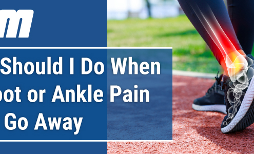 https://orthosportsmed.com/wp-content/uploads/2024/04/Blog-header-image-what-should-i-do-when-my-foot-or-ankle-pain-wont-go-away-OSM-Oregon.jpg
300
833
orthosportsmed
https://orthosportsmed.com/wp-content/uploads/2015/01/osm-header-vs7.png
orthosportsmed2024-04-16 12:00:282024-04-01 16:00:22What Should I Do When My Foot or Ankle Pain Won’t Go Away?
https://orthosportsmed.com/wp-content/uploads/2024/04/Blog-header-image-what-should-i-do-when-my-foot-or-ankle-pain-wont-go-away-OSM-Oregon.jpg
300
833
orthosportsmed
https://orthosportsmed.com/wp-content/uploads/2015/01/osm-header-vs7.png
orthosportsmed2024-04-16 12:00:282024-04-01 16:00:22What Should I Do When My Foot or Ankle Pain Won’t Go Away? https://orthosportsmed.com/wp-content/uploads/2024/02/Blog-header-image-why-go-see-a-podiatrist-orthopedic-surgery-outcomes-OSM-Oregon.jpg
300
833
orthosportsmed
https://orthosportsmed.com/wp-content/uploads/2015/01/osm-header-vs7.png
orthosportsmed2024-02-02 12:00:342024-02-05 09:33:22Why Go See a Podiatrist?
https://orthosportsmed.com/wp-content/uploads/2024/02/Blog-header-image-why-go-see-a-podiatrist-orthopedic-surgery-outcomes-OSM-Oregon.jpg
300
833
orthosportsmed
https://orthosportsmed.com/wp-content/uploads/2015/01/osm-header-vs7.png
orthosportsmed2024-02-02 12:00:342024-02-05 09:33:22Why Go See a Podiatrist? https://orthosportsmed.com/wp-content/uploads/2023/10/Blog-header-image-what-is-minimally-invasive-foot-surgery-OSM-Oregon.jpg
300
833
orthosportsmed
https://orthosportsmed.com/wp-content/uploads/2015/01/osm-header-vs7.png
orthosportsmed2023-10-10 12:00:242023-10-02 13:36:14What is Minimally Invasive Foot Surgery?
https://orthosportsmed.com/wp-content/uploads/2023/10/Blog-header-image-what-is-minimally-invasive-foot-surgery-OSM-Oregon.jpg
300
833
orthosportsmed
https://orthosportsmed.com/wp-content/uploads/2015/01/osm-header-vs7.png
orthosportsmed2023-10-10 12:00:242023-10-02 13:36:14What is Minimally Invasive Foot Surgery?