Spine Structure and Function

Article featured on the Cleveland Clinic
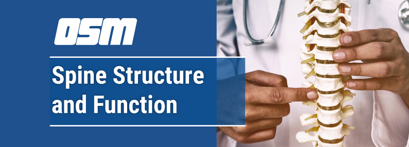
Key parts of your spine include vertebrae (bones), disks, nerves and the spinal cord. The spine supports your body and helps you walk, twist and move. The disks that cushion vertebrae may compress with age or injury, leading to a herniated disk. Exercises can strengthen the core muscles that support the spine and prevent back injuries and back pain.

What is the spine?

Your spine, or backbone, is your body’s central support structure. It connects different parts of your musculoskeletal system. Your spine helps you sit, stand, walk, twist and bend. Back injuries, spinal cord conditions and other problems can damage the spine and cause back pain.

Cleveland Clinic is a non-profit academic medical center. Advertising on our site helps support our mission. We do not endorse non-Cleveland Clinic products or services.

What are the parts of the spine?

A healthy spine has three natural curves that make an S-shape. These curves absorb shocks to your body and protect your spine from injury. Many different parts make up your spine:

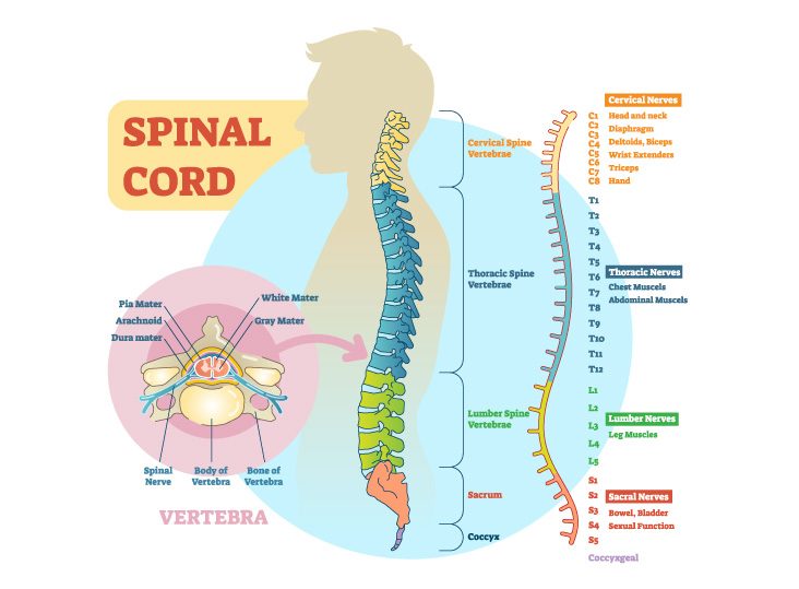
  • Vertebrae: The spine has 33 stacked vertebrae (small bones) that form the spinal canal. The spinal canal is a tunnel that houses the spinal cord and nerves, protecting them from injury. Most vertebrae move to allow for a range of motion. The lowest vertebrae (sacrum and coccyx) are fused together and don’t move.
  • Facet joints: These spinal joints have cartilage (a slippery connective tissue) that allows vertebrae to slide against each other. Facet joints let you twist and turn, and they provide flexibility and stability. These joints can develop arthritis and cause back pain or neck pain.
  • Intervertebral disks: These flat, round cushions sit between the vertebrae and act as the spine’s shock absorbers. Each disk has a soft, gel-like center (the nucleus pulposus) surrounded by a flexible outer ring (the annulus). Intervertebral disks are under constant pressure. A herniated disk can tear, allowing some of the nucleus’ gel substance to leak out. Herniated disks (also called bulging, slipped or ruptured disks) can be painful.
  • Spinal cord and nerves: The spinal cord is a column of nerves that travels through the spinal canal. The cord extends from the skull to the lower back. Thirty-one pairs of nerves branch out through vertebral openings (the neural foramen). These nerves carry messages between the brain and muscles.
  • Soft tissues: Ligaments connect the vertebrae to hold the spine in position. Muscles support the back and help you move. Tendons connect muscles to bone and aid movement.

What are the spine segments?

The 33 vertebrae make up five distinct spine segments. Starting at the neck and going down toward your buttocks (rear end), these segments include:

  • Cervical (neck): The top part of the spine has seven vertebrae (C1 to C7). These neck vertebrae allow you to turn, tilt and nod your head. The cervical spine makes an inward C-shape called a lordotic curve.
  • Thoracic (middle back): The chest or thoracic part of the spine has 12 vertebrae (T1 to T12). Your ribs attach to the thoracic spine. This section of the spine bends out slightly to make a backward C-shape called the kyphotic curve.
  • Lumbar (lower back): Five vertebrae (L1 to L5) make up the lower part of the spine. Your lumbar spine supports the upper parts of the spine. It connects to the pelvis and bears most of your body’s weight, as well as the stress of lifting and carrying items. Many back problems occur in the lumbar spine. The lumbar spine bends inward to create a C-shaped lordotic curve.
  • Sacrum: This triangle-shaped bone connects to the hips. The five sacral vertebrae (S1 to S5) fuse as a baby develops in the womb, which means they don’t move. The sacrum and hip bones form a ring called the pelvic girdle.
  • Coccyx (tailbone): Four fused vertebrae make up this small piece of bone found at the bottom of the spine. Pelvic floor muscles and ligaments attach to the coccyx.

What conditions and disorders affect the spine?

Up to 80% of Americans experience back pain at some point. Vertebrae and disks can wear down with age, causing pain. Other conditions that affect spine health include:

  • Arthritic conditions, such as ankylosing spondylitis (AS).
  • Back strains and sprains.
  • Birth defects such as spina bifida.
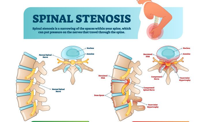
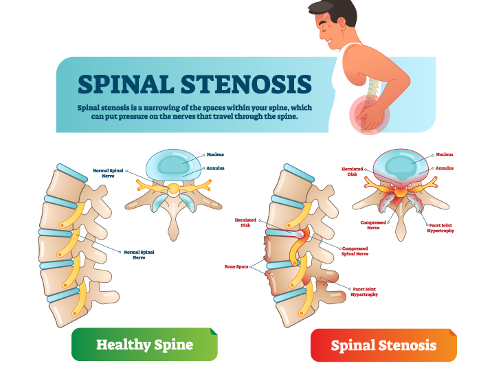
  • Bone spurs (jagged edges on vertebrae that put pressure on the spinal cord and nerves).
  • Curvatures of the spine (scoliosis and kyphosis).
  • Neuromuscular diseases, such as amyotrophic lateral sclerosis (ALS).
  • Nerve injuries, including spinal stenosis, sciatica and pinched nerves.
  • Osteoporosis (weak bones).
  • Spinal cord injuries, including spinal fractures, herniated disks and paralysis.
  • Spine tumors and cancer.
  • Spine infections like meningitis and osteomyelitis.

How can I keep my spine healthy?

Strong back muscles can protect your spine and prevent back problems. Try to do back-strengthening and stretching exercises at least twice a week. Exercises like planks strengthen the core (abdominal, side and back muscles) to give your spine more support. Other protective measures include:

  • Bend your knees and keep your back straight when lifting items.
  • Lose weight, if needed (excess weight strains your back).
  • Maintain good posture.

When should I call the doctor?

You should call your healthcare provider if you experience:

  • Back pain with fever.
  • Bowel or bladder control issues.
  • Leg weakness or pain that moves from your back down your legs.
  • Pain that worsens, causes nausea or sleeplessness or interferes with daily activities.

The Orthopedic & Sports Medicine Center of Oregon is an award-winning, board-certified orthopedic group located in downtown Portland Oregon. We utilize both surgical and nonsurgical means to treat musculoskeletal trauma, spine diseases, sports injuries, degenerative diseases, infections, tumors and congenital disorders.

Our mission is to return our patients back to pain-free mobility and full strength as quickly and painlessly as possible using both surgical and non-surgical orthopedic procedures.

Our expert physicians provide leading-edge, comprehensive care in the diagnosis and treatment of orthopedic conditions, including total joint replacement and sports medicine. We apply the latest state-of-the-art techniques in order to return our patients to their active lifestyle.

If you’re looking for compassionate, expert orthopedic surgeons in Portland Oregon, contact OSM today.

Phone:
503-224-8399

Address
1515 NW 18th Ave, 3rd Floor
Portland, OR 97209

Hours
Monday–Friday

Cervical Disc Surgery: Disc Replacement or Fusion?

Article featured on WebMD, medically reviewed by Sabrina Felson, MD

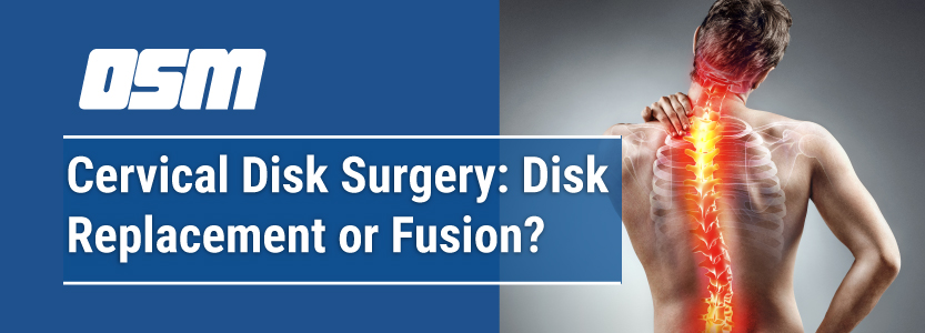
The vast majority of people — more than 90% — with pain from cervical disc disease will get better on their own over time with simple, conservative treatments. Surgery, however, may help if other treatments fail or if symptoms worsen to the point that weakness in your arms and or legs develop. This is called a cervical myelopathy and surgery is recommended.

Cervical disc disease is caused by an abnormality in one or more discs — the cushions — that lie between the neck bones (vertebrae). When a disc is damaged — due to degenerative disc disease (or DDD) or an unknown cause — it can lead to neck pain from inflammation or muscle spasm. In severe cases, pain and numbness can occur in the arms from pressure on the cervical nerve roots or spinal cord.

Surgery for cervical disc disease typically involves removing the disc that is pinching the nerve or pressing on the spinal cord. This surgery is called a discectomy. Depending on where the disc is located, the surgeon can remove it through a small incision either in the front (anterior discectomy) or back (posterior discectomy) of the neck while you are under anesthesia. A similar technique, microdiscectomy, removes the disc through a smaller incision using a microscope or other magnifying device.

Often, a procedure is performed to close the space that’s left when the disc is removed and restore the spine to its original length. Patients have two options:

  • Artificial cervical disc replacement
  • Cervical fusion

In 2007, the FDA approved the first artificial disc, the Prestige Cervical disc, which looks and moves much like the real thing but is made of metal. Since then, several artificial cervical discs have been developed and approved. Ongoing research has shown that the artificial disc can improve neck and arm pain as safely and effectively as cervical fusion while allowing for range of motion that is as good or better than with cervical fusion. People who get the artificial disc are often able to return to work more quickly as well. The surgery to replace the disc, however, does take longer and can lead to more blood loss than with cervical fusion. It’s also not known how the artificial discs will last over time. People who get an artificial disc can always opt for cervical fusion later. But if a patient has cervical fusion first, it’s not possible to later put an artificial disc in the same spot.


The Orthopedic & Sports Medicine Center of Oregon is an award-winning, board-certified orthopedic group located in downtown Portland Oregon. We utilize both surgical and nonsurgical means to treat musculoskeletal trauma, spine diseases, sports injuries, degenerative diseases, infections, tumors and congenital disorders.

Our mission is to return our patients back to pain-free mobility and full strength as quickly and painlessly as possible using both surgical and non-surgical orthopedic procedures.

Our expert physicians provide leading-edge, comprehensive care in the diagnosis and treatment of orthopedic conditions, including total joint replacement and sports medicine. We apply the latest state-of-the-art techniques in order to return our patients to their active lifestyle.

If you’re looking for compassionate, expert orthopedic surgeons in Portland Oregon, contact OSM today.

Phone:
503-224-8399

Address
1515 NW 18th Ave, 3rd Floor
Portland, OR 97209

Hours
Monday–Friday

8:00am – 4:30pm

Symptoms & Solutions: Spinal Stenosis

Article featured on Arkansas Surgical Hospital

What is Spinal Stenosis?

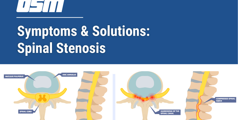
Spinal stenosis is a deterioration of vertebrae that causes narrowing of the openings of the spinal column.  Over time, this puts pressure on the nerves inside the spinal column.  In most cases, individuals develop spinal stenosis in either the neck (called cervical stenosis) or the lower back (called lumbar stenosis).

What Causes Spinal Stenosis?

The most common cause of spinal stenosis is osteoarthritis that causes bone spurs.  These spurs can impinge on the spinal cord or pinch one or more nerves emanating from the spinal column.  Other causes of spinal stenosis include pressure on the spinal cord or nerves from herniated discs, tumors, thickened or inflamed ligaments, or trauma that dislocates or breaks vertebrae.

Types of Spinal Stenosis & Their Symptoms

Some people develop mild cases of spinal stenosis without experiencing any symptoms.  However, if the deterioration continues over time, a variety of symptoms can develop.  Symptoms are most common in individuals over 60 years of age and tend to become more severe with age due to wear and tear on the spinal vertebrae. Symptoms typically vary based on the location of the stenosis in the spine.

Lumbar Spinal Stenosis

Spinal stenosis is more common in the lower back than in the neck.  This type of spinal stenosis is called lumbar stenosis.  The most common symptom of lumbar stenosis is neurogenic claudication, which is leg pain that comes and goes due to pinched nerves in the spinal cord.  It can cause weakness or cramps in the legs that can be severe.  It is more troublesome when standing or walking for long periods, and it may become less severe or go away when you sit or bend forward.

Numbness or a pins-and-needles sensation in one or both legs is another symptom.  The sensation is similar to when your foot falls asleep, but is more frequent or may last longer.  Weakness in the feet or legs can also indicate lumbar stenosis.  Gait issues such as foot drop may arise from weakness in the calves or quadriceps.  Foot drop is the inability to raise the front of the foot properly due to weakness.  Instead, the foot drops forward, often dragging on the ground or causing the person to trip.

When the pain is in one leg and accompanied by weakness, it may be referred to as sciatica, depending on which nerve is compressed.  Some individuals may feel pain in the buttocks and lower back as well.

Cervical Spinal Stenosis

The narrowing of the spinal column that causes pressure on the spinal cord in the neck is called cervical stenosis.  People with cervical stenosis can develop problems with their gait and keeping their balance, which affects their mobility.  The gait problems arise from the compression of the spinal cord rather than a pinched nerve.  This condition progresses over time, with the individual falling more frequently as the compression worsens.

Pain in the shoulder, arm, or neck may also be a sign of cervical stenosis, particularly if the pain is shock-like or is accompanied by a burning sensation.  Some people experience numbness or a pins-and-needles tingling sensation in one or both hands.  This is sometimes accompanied by weakness and loss of fine motor skills.  Over time, it becomes increasingly harder to fasten buttons, use a pen, or perform simple tasks that require small movements.

In exceedingly rare cases, individuals with severe spinal stenosis may experience incontinence, severe weakness in the legs, or loss of feeling in the genitals and inner thighs.  If this happens, contact emergency medical help immediately.  For most people, spinal stenosis is diagnosed and treated before it progresses to this point.

Diagnosing Spinal Stenosis

Your doctor will initially diagnose cervical or lumbar stenosis based on your symptoms, medical history, risk factors (such as age and injury), and a complete physical exam.  To confirm the diagnosis, they will use imaging procedures to determine the cause of your symptoms.  These may include x-rays of the spinal column, MRIs, and CT scans.  These techniques can reveal bone spurs, herniated discs, tumors, and areas where there is pressure on the spinal cord or nerves.

Conservative Treatments for Spinal Stenosis

There are several treatment options for mild to moderate spinal stenosis.  Your doctor can help determine the best treatment (or combination of treatments) for you based on the severity of your condition and your pain level.

Conservative treatments for mild to moderate spinal stenosis may include:

  • Pain relievers such as naproxen, ibuprofen, or acetaminophen for short-term relief
  • Antidepressants to minimize chronic pain and any resulting minor depression
  • Prescription opioids such as hydrocodone (used sparingly for short periods of time)
  • Physical therapy to improve muscle tone and strength, maintain spinal stability, and improve balance
  • Steroid injections to reduce the inflammation of irritated nerves

While these methods don’t correct spinal stenosis and may not work for everyone, they are typically done to reduce swelling, pain, and other symptoms.  Surgery is the last resort after other options for alleviating the symptoms of spinal stenosis have proven ineffective.

Surgical Solutions for Spinal Stenosis

When the symptoms of spinal stenosis become moderate or severe, surgery may be the only option.  This is particularly important when neurological issues, pain, or mobility problems have developed.  The goal of spinal stenosis surgery is to relieve pressure on the nerves or spinal cord so they can heal and return to proper functioning.

Laminectomy

The most common surgical treatment for either cervical or lumbar stenosis is a laminectomy.  During this procedure, two vertebral laminae—which form the “roof” of the spinal canal—and the bony area connecting them are removed, relieving pressure on the spinal cord.  Bone grafts or instrumentation are added to stabilize the site and protect the spinal cord.  Depending on the severity of the stenosis, fusion of the surrounding vertebrae may be performed for added stability.

Transforaminotomy

Transforaminotomy is generally for less severe cases of lumbar stenosis.  This option is not as involved as a laminectomy and is reserved for situations when the stenosis is restricted to a small area of the lumbar spine.  Transforaminotomy involves the widening of the bony openings between affected vertebrae, including the removal of any bone spurs that have developed.  Your surgeon may also remove damaged soft tissue or herniated discs.

Discectomy

If spinal stenosis is caused by a degenerated or herniated disc pressing on a nerve or the spinal cord, discectomy surgery may be done to remove all or part of the affected disc.  Partial discectomies are typically performed for lumbar stenosis, while complete discectomies are more common for cervical stenosis.  Your surgeon may fuse the surrounding vertebrae if needed.

Any of these surgical procedures may include minimally invasive options, depending on the severity of the spinal stenosis.  After surgery, appropriate care must be taken to allow the area to heal.  In some cases, patients will need physical therapy to ensure proper mobility following surgery.


The Orthopedic & Sports Medicine Center of Oregon is an award-winning, board-certified orthopedic group located in downtown Portland Oregon. We utilize both surgical and nonsurgical means to treat musculoskeletal trauma, spine diseases, sports injuries, degenerative diseases, infections, tumors and congenital disorders.

Our mission is to return our patients back to pain-free mobility and full strength as quickly and painlessly as possible using both surgical and non-surgical orthopedic procedures.

Our expert physicians provide leading-edge, comprehensive care in the diagnosis and treatment of orthopedic conditions, including total joint replacement and sports medicine. We apply the latest state-of-the-art techniques in order to return our patients to their active lifestyle.

If you’re looking for compassionate, expert orthopedic surgeons in Portland Oregon, contact OSM today.

Phone:
503-224-8399

Address
1515 NW 18th Ave, 3rd Floor
Portland, OR 97209

Hours
Monday–Friday
8:00am – 4:30pm

What Is Spinal Fusion?

From WebMD


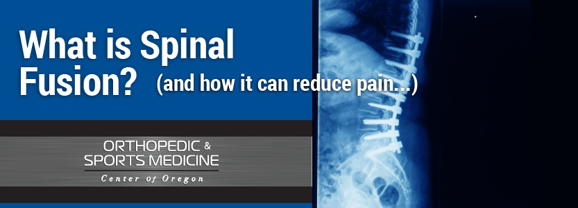
Spinal fusion is surgery to join two or more vertebrae into one single structure. The goal is to stop movement between the two bones and prevent back pain. Once they’re fused, they no longer move like they used to. This keeps you from stretching nearby nerves, ligaments, and muscles that may have caused discomfort.

Spinal fusion involves techniques designed to mimic the normal healing process of broken bones. During spinal fusion, your surgeon places bone or a bonelike material within the space between two spinal vertebrae. Metal plates, screws and rods may be used to hold the vertebrae together, so they can heal into one solid unit. Read more

What is Spinal Stenosis?

From WebMD

Cervical Spinal Stenosis

Spinal stenosis is a condition, mostly in adults 50 and older, in which your spinal canal starts to narrow. This can cause pain and other problems.

Your spine is made up of a series of connected bones (or “vertebrae”) and shock-absorbing discs. It protects your spinal cord, a key part of the central nervous system that connects the brain to the body. The cord rests in the canal formed by your vertebrae.

For most people, the stenosis results from changes because of arthritis. The spinal canal may narrow. The open spaces between the vertebrae may start to get smaller. The tightness can pinch the spinal cord or the nerves around it, causing pain, tingling, or numbness in your legs, arms, or torso. Read more

How The Spinal Cord Works

Article shared from ChristopherReeve.org

What is the central nervous system?

The central nervous system (CNS) controls most functions of the body and mind. It consists of two parts: the brain and the spinal cord.

The brain is the center of our thoughts, the interpreter of our external environment, and the origin of control over body movement. Like a central computer, it interprets information from our eyes (sight), ears (sound), nose (smell), tongue (taste), and skin (touch), as well as from internal organs such as the stomach.
Read more