
 https://orthosportsmed.com/wp-content/uploads/2023/06/Blog-header-image-treating-hand-arthritis-without-surgery-OSM-Oregon.jpg
300
833
orthosportsmed
https://orthosportsmed.com/wp-content/uploads/2015/01/osm-header-vs7.png
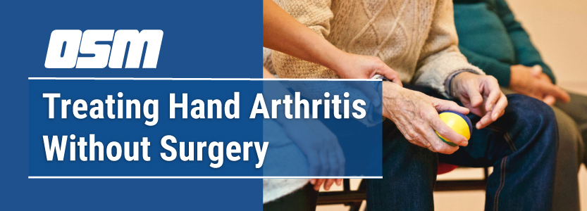
orthosportsmed2023-06-20 12:00:342023-07-27 21:53:24Treating Hand Arthritis Without Surgery
https://orthosportsmed.com/wp-content/uploads/2023/06/Blog-header-image-treating-hand-arthritis-without-surgery-OSM-Oregon.jpg
300
833
orthosportsmed
https://orthosportsmed.com/wp-content/uploads/2015/01/osm-header-vs7.png
orthosportsmed2023-06-20 12:00:342023-07-27 21:53:24Treating Hand Arthritis Without Surgery https://orthosportsmed.com/wp-content/uploads/2023/03/Blog-header-image-how-to-manage-and-prevent-arthritis-in-the-hand-OSM-Oregon.jpg
300
833
orthosportsmed
https://orthosportsmed.com/wp-content/uploads/2015/01/osm-header-vs7.png
orthosportsmed2023-03-29 12:00:002023-03-01 02:16:58How to Manage & Prevent Arthritis in the Hands
https://orthosportsmed.com/wp-content/uploads/2023/03/Blog-header-image-how-to-manage-and-prevent-arthritis-in-the-hand-OSM-Oregon.jpg
300
833
orthosportsmed
https://orthosportsmed.com/wp-content/uploads/2015/01/osm-header-vs7.png
orthosportsmed2023-03-29 12:00:002023-03-01 02:16:58How to Manage & Prevent Arthritis in the Hands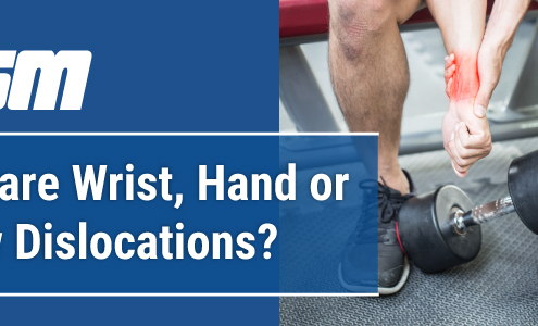 https://orthosportsmed.com/wp-content/uploads/2023/03/Blog-header-image-what-are-wrist-hand-or-elbow-dislocations-OSM-Oregon.jpg
300
833
orthosportsmed
https://orthosportsmed.com/wp-content/uploads/2015/01/osm-header-vs7.png
orthosportsmed2023-03-07 12:00:532023-03-01 02:06:08What are Wrist, Hand or Elbow Dislocations?
https://orthosportsmed.com/wp-content/uploads/2023/03/Blog-header-image-what-are-wrist-hand-or-elbow-dislocations-OSM-Oregon.jpg
300
833
orthosportsmed
https://orthosportsmed.com/wp-content/uploads/2015/01/osm-header-vs7.png
orthosportsmed2023-03-07 12:00:532023-03-01 02:06:08What are Wrist, Hand or Elbow Dislocations?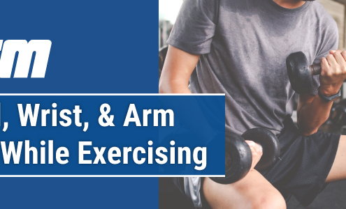 https://orthosportsmed.com/wp-content/uploads/2022/10/Blog-header-image-hand-wrist-and-arm-pain-while-exercising-OSM-Oregon.jpg
300
833
orthosportsmed
https://orthosportsmed.com/wp-content/uploads/2015/01/osm-header-vs7.png
orthosportsmed2022-10-25 12:00:012022-10-05 12:12:20Hand, Wrist, And Arm Pain While Exercising
https://orthosportsmed.com/wp-content/uploads/2022/10/Blog-header-image-hand-wrist-and-arm-pain-while-exercising-OSM-Oregon.jpg
300
833
orthosportsmed
https://orthosportsmed.com/wp-content/uploads/2015/01/osm-header-vs7.png
orthosportsmed2022-10-25 12:00:012022-10-05 12:12:20Hand, Wrist, And Arm Pain While Exercising https://orthosportsmed.com/wp-content/uploads/2022/07/Blog-header-image-5-causes-of-thumb-pain-OSM-Oregon-1.jpg
300
833
orthosportsmed
https://orthosportsmed.com/wp-content/uploads/2015/01/osm-header-vs7.png
orthosportsmed2022-07-19 12:00:392022-06-30 20:15:175 Causes of Thumb Pain
https://orthosportsmed.com/wp-content/uploads/2022/07/Blog-header-image-5-causes-of-thumb-pain-OSM-Oregon-1.jpg
300
833
orthosportsmed
https://orthosportsmed.com/wp-content/uploads/2015/01/osm-header-vs7.png
orthosportsmed2022-07-19 12:00:392022-06-30 20:15:175 Causes of Thumb Pain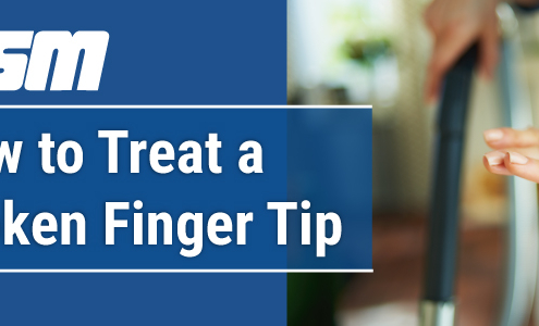 https://orthosportsmed.com/wp-content/uploads/2022/05/Blog-header-image-how-to-treat-a-broken-finger-tip-OSM-Oregon.jpg
300
833
orthosportsmed
https://orthosportsmed.com/wp-content/uploads/2015/01/osm-header-vs7.png
orthosportsmed2022-05-12 12:00:472022-04-22 18:37:25How to Treat a Broken Finger Tip
https://orthosportsmed.com/wp-content/uploads/2022/05/Blog-header-image-how-to-treat-a-broken-finger-tip-OSM-Oregon.jpg
300
833
orthosportsmed
https://orthosportsmed.com/wp-content/uploads/2015/01/osm-header-vs7.png
orthosportsmed2022-05-12 12:00:472022-04-22 18:37:25How to Treat a Broken Finger Tip https://orthosportsmed.com/wp-content/uploads/2021/11/Blog-header-image-hand-pain-from-carpal-tunnel-or-something-else-OSM-Oregon.jpg
300
833
orthosportsmed
https://orthosportsmed.com/wp-content/uploads/2015/01/osm-header-vs7.png
orthosportsmed2021-11-23 12:00:442022-03-01 14:28:11Is My Hand Pain from Carpal Tunnel Syndrome or Something Else?
https://orthosportsmed.com/wp-content/uploads/2021/11/Blog-header-image-hand-pain-from-carpal-tunnel-or-something-else-OSM-Oregon.jpg
300
833
orthosportsmed
https://orthosportsmed.com/wp-content/uploads/2015/01/osm-header-vs7.png
orthosportsmed2021-11-23 12:00:442022-03-01 14:28:11Is My Hand Pain from Carpal Tunnel Syndrome or Something Else? https://orthosportsmed.com/wp-content/uploads/2021/07/Blog-header-image-what-to-do-hand-wrist-pain-OSM-Oregon.jpg
300
833
orthosportsmed
https://orthosportsmed.com/wp-content/uploads/2015/01/osm-header-vs7.png
orthosportsmed2021-07-06 12:00:162021-06-30 18:19:46What to Do for Hand and Wrist Pain
https://orthosportsmed.com/wp-content/uploads/2021/07/Blog-header-image-what-to-do-hand-wrist-pain-OSM-Oregon.jpg
300
833
orthosportsmed
https://orthosportsmed.com/wp-content/uploads/2015/01/osm-header-vs7.png
orthosportsmed2021-07-06 12:00:162021-06-30 18:19:46What to Do for Hand and Wrist Pain https://orthosportsmed.com/wp-content/uploads/2020/06/Arthritis-of-the-Hand.jpg
320
845
orthosportsmed
https://orthosportsmed.com/wp-content/uploads/2015/01/osm-header-vs7.png
orthosportsmed2020-06-20 04:32:002020-07-11 15:10:13Arthritis of the Hand
https://orthosportsmed.com/wp-content/uploads/2020/06/Arthritis-of-the-Hand.jpg
320
845
orthosportsmed
https://orthosportsmed.com/wp-content/uploads/2015/01/osm-header-vs7.png
orthosportsmed2020-06-20 04:32:002020-07-11 15:10:13Arthritis of the Hand https://orthosportsmed.com/wp-content/uploads/2020/01/What-Is-Dupuytrens-Disease-and-How-Is-It-Treated.jpg
320
845
orthosportsmed
https://orthosportsmed.com/wp-content/uploads/2015/01/osm-header-vs7.png
orthosportsmed2020-01-11 22:05:442020-01-11 22:05:44What Is Dupuytren’s Disease and How Is It Treated?
https://orthosportsmed.com/wp-content/uploads/2020/01/What-Is-Dupuytrens-Disease-and-How-Is-It-Treated.jpg
320
845
orthosportsmed
https://orthosportsmed.com/wp-content/uploads/2015/01/osm-header-vs7.png
orthosportsmed2020-01-11 22:05:442020-01-11 22:05:44What Is Dupuytren’s Disease and How Is It Treated?