
 https://orthosportsmed.com/wp-content/uploads/2026/01/Blog-header-image-hand-arthritis-symptoms-and-treatment-Oregon-OSM.png
320
846
orthosportsmed
https://orthosportsmed.com/wp-content/uploads/2015/01/osm-header-vs7.png
orthosportsmed2026-01-15 12:00:062026-01-03 10:24:34Hand Arthritis Symptoms and Treatment
https://orthosportsmed.com/wp-content/uploads/2026/01/Blog-header-image-hand-arthritis-symptoms-and-treatment-Oregon-OSM.png
320
846
orthosportsmed
https://orthosportsmed.com/wp-content/uploads/2015/01/osm-header-vs7.png
orthosportsmed2026-01-15 12:00:062026-01-03 10:24:34Hand Arthritis Symptoms and Treatment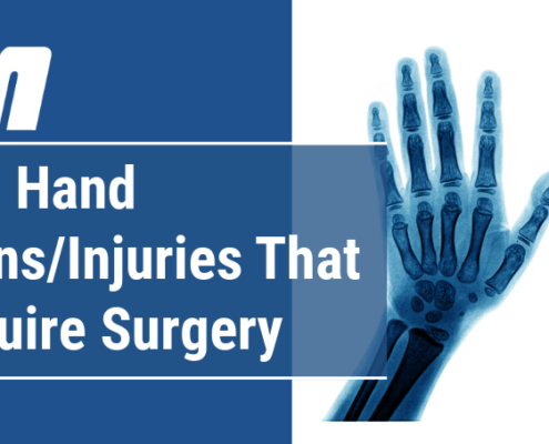 https://orthosportsmed.com/wp-content/uploads/2025/01/Blog-header-image-common-hand-injuries-conditional-that-may-require-surgery-Oregon-OSM.png
454
1199
orthosportsmed
https://orthosportsmed.com/wp-content/uploads/2015/01/osm-header-vs7.png
orthosportsmed2025-01-23 12:00:132025-01-05 14:23:26Common Hand Conditions or Injuries That May Require Surgery
https://orthosportsmed.com/wp-content/uploads/2025/01/Blog-header-image-common-hand-injuries-conditional-that-may-require-surgery-Oregon-OSM.png
454
1199
orthosportsmed
https://orthosportsmed.com/wp-content/uploads/2015/01/osm-header-vs7.png
orthosportsmed2025-01-23 12:00:132025-01-05 14:23:26Common Hand Conditions or Injuries That May Require Surgery https://orthosportsmed.com/wp-content/uploads/2020/01/What-Is-Dupuytrens-Disease-and-How-Is-It-Treated.jpg
320
845
orthosportsmed
https://orthosportsmed.com/wp-content/uploads/2015/01/osm-header-vs7.png
orthosportsmed2020-01-11 22:05:442020-01-11 22:05:44What Is Dupuytren’s Disease and How Is It Treated?
https://orthosportsmed.com/wp-content/uploads/2020/01/What-Is-Dupuytrens-Disease-and-How-Is-It-Treated.jpg
320
845
orthosportsmed
https://orthosportsmed.com/wp-content/uploads/2015/01/osm-header-vs7.png
orthosportsmed2020-01-11 22:05:442020-01-11 22:05:44What Is Dupuytren’s Disease and How Is It Treated? https://orthosportsmed.com/wp-content/uploads/2018/06/Kienbocks-Disease.jpg
320
845
orthosportsmed
https://orthosportsmed.com/wp-content/uploads/2015/01/osm-header-vs7.png
orthosportsmed2018-06-14 03:38:212018-06-14 08:06:21What Is Kienböck’s Disease?
https://orthosportsmed.com/wp-content/uploads/2018/06/Kienbocks-Disease.jpg
320
845
orthosportsmed
https://orthosportsmed.com/wp-content/uploads/2015/01/osm-header-vs7.png
orthosportsmed2018-06-14 03:38:212018-06-14 08:06:21What Is Kienböck’s Disease?