What to Know About Forearm Muscles

Article featured on WebMD

Many muscles make up the forearm, extending from your elbow joint to your hand. The ulna and radius bones form a rotational joint that allows your forearm to turn the palm of your hand either up or down. Two large arteries, also known as the ulna and radius, run the length of the forearm and branch into smaller arteries that service your forearm’s musculature.

The bones in your forearm are prone to being broken because people often instinctually extend their forearm trying to break a fall or protect their face, which could lead to a fracture. The muscles in your forearm that allow you to bring about different movements can be categorized as anterior and posterior.

These are the muscles that can be found in your forearm:

The Anterior Compartment 

The anterior superficial layer contains four muscles that originate from the medial epicondyle. The pronator teres muscle attaches to the shaft of the radius and is the most medial of the muscles in this layer. Its primary action is the pronation of the forearm. The flexor carpi radialis contributes to abduction and attaches to the base of metacarpals II and III.

Connecting to the flexor retinaculum and acting to flex at the wrist, the palmaris longus allows you to wave at a friend or say goodbye to a loved one. About 15% of the population does not have this muscle, though.

The flexor carpi ulnar is the most lateral of the muscles in the superficial layer, responsible for flexion and abduction at the wrist. It attaches the hand to the pisiform bone and base of the 5th metacarpal. This muscle allows you to move your wrist back and forth.

The Intermediate Layer 

Only one muscle makes up the intermediate layer, which originates from the medial condyle of the humerus and the radius. The flexor digitorum superficialis (FDS) lies between the deep and superficial muscle layers and splits into four tendons that attach to the middle phalanx of a finger. At the proximal interphalangeal joints (PIPJs) and the metacarpophalangeal joints (MCPJs), the FDS flexes the fingers and contributes to wrist flexion.

The Deep Layer 

The flexor digitorum profundus (FDP) splits into four tendons and originates at the ulna. This muscle attaches to the distal phalanx of each finger and allows flexion of the metacarpophalangeal joints and distal interphalangeal joints. Bending your ring, middle, index, and pinkie fingers is possible because of this muscle. The flexor pollicis longus extends laterally to the flexor digitorum profundus muscle.

This muscle attaches to the distal phalanx of the thumb and originates from the radius. It stretches laterally to the FDP and allows you to bend your thumb. A square-shaped muscle found in the FDL and FDP, the pronator quadratus attaches to the radius and originates from the ulna. The pronator quadratus allows you to pronate your forearm and is innervated by the median nerve.

Posterior Compartment 

The radial nerve innervates all the muscles in this compartment; it contains more muscles than the anterior compartment and can be split into the superficial and deep layers. Eight muscles are in the superficial layer, and five are in the deep compartment. Four of the five muscles in the deep layer act on your thumb and index finger.
The Superficial Layer The brachioradialis muscle attaches to the distal radius and originates from the lateral humerus. It allows you to flex your elbow and lift a glass of water to your mouth. Contributing to wrist abduction, the extensor carpi radialis longus muscle acts to extend the wrist. It attaches to the metacarpal III and originates from the lateral aspect of the humerus. The extensor digitorum splits into four tendons and connects to the distal phalanx of each of your fingers. Originating from the lateral epicondyle of the humerus, this muscle acts to extend your fingers at the PIJs and MCPJs.The extensor digiti minimi originates from the lateral epicondyle of the humerus and acts to extend the little finger. The extensor carpi ulnaris attaches to metacarpal V and originates from the lateral epicondyle of the humerus. It contributes to wrist abduction and acts to extend at the wrist. The anconeus is situated superior to the other muscles in the superficial layer. The anconeus attaches to the olecranon and originates from the lateral epicondyle. It acts to extend at your elbow joint.

The Deep Layer

The supinator muscle can be found in the deep layer and originates from the lateral epicondylitis of the humerus and the ulna. The deep radial nerve innervates this muscle.

The extensor pollicis brevis allows you to make a thumbs-up signal. It attaches to the proximal phalanx of the thumb and originates from the posterior radius. Your extensor pollicis longus muscle acts to extend the thumb and is attached to the distal phalanx of the thumb. The extensor indicis attaches to the distal phalanx of your index finger and also acts to extend it; this muscle originates from the interosseous membrane and the ulna.

Your abductor pollicis longus muscle attaches to metacarpal I and abducts the thumb. It sits between the radius and ulna and originates from the interosseous membrane.

From taking a heavy box up a flight of stairs to playing basketball, your forearm muscles are used in your daily life, and strengthening these muscles can also help increase your grip strength. A firm grip can help you in many ways in your everyday life.

Here are some exercises you can try to strengthen your arm muscles:

Find weighted objects like tires to carry and walk for as long as possible. Set it down and then pick it back up. Repeat this until you get tired. If you have access to a sturdy bar, try pull-ups or chin-ups. Other activities like gardening are an excellent way to keep your body in motion and strengthen your arms and hands. Pulling weeds and hammering nails can also help you achieve stronger forearms.

If you go to the gym, grab a couple of dumbbells, and hold them out in front of you until you are exhausted. Finger curls, dumbbell reverse curls, barbell reverse bicep curls, dumbbell wrist extensions, and hammer curls are great exercises you can try to strengthen your forearms.


The Orthopedic & Sports Medicine Center of Oregon is an award-winning, board-certified orthopedic group located in downtown Portland Oregon. We utilize both surgical and nonsurgical means to treat musculoskeletal trauma, spine diseases, foot and ankle conditions, sports injuries, degenerative diseases, infections, tumors and congenital disorders.

Our mission is to return our patients back to pain-free mobility and full strength as quickly and painlessly as possible using both surgical and non-surgical orthopedic procedures.

Our expert physicians provide leading-edge, comprehensive care in the diagnosis and treatment of orthopedic conditions, including total joint replacement and sports medicine. We apply the latest state-of-the-art techniques in order to return our patients to their active lifestyle.

If you’re looking for compassionate, expert orthopedic and podiatric surgeons in Portland Oregon, contact OSM today.

Phone:

503-224-8399

Address
1515 NW 18th Ave, 3rd Floor
Portland, OR 97209

Hours
Monday–Friday
8:00am – 4:30pm

Baseball Season is Here: Watch out for UCL Tears


The Orthopedic & Sports Medicine Center of Oregon is an award-winning, board-certified orthopedic group located in downtown Portland Oregon. We utilize both surgical and nonsurgical means to treat musculoskeletal trauma, spine diseases, foot and ankle conditions, sports injuries, degenerative diseases, infections, tumors and congenital disorders.

Our mission is to return our patients back to pain-free mobility and full strength as quickly and painlessly as possible using both surgical and non-surgical orthopedic procedures.

Our expert physicians provide leading-edge, comprehensive care in the diagnosis and treatment of orthopedic conditions, including total joint replacement and sports medicine. We apply the latest state-of-the-art techniques in order to return our patients to their active lifestyle.

If you’re looking for compassionate, expert orthopedic and podiatric surgeons in Portland Oregon, contact OSM today.

Phone:

503-224-8399

Address
1515 NW 18th Ave, 3rd Floor
Portland, OR 97209

Hours
Monday–Friday
8:00am – 4:30pm

What are Wrist, Hand or Elbow Dislocations?

Article featured on Mercy Health

What are hand, wrist or elbow dislocations?

Hand dislocations occur when one of the eight carpal bones (bones located at the base of the hand) fall out of the joint to cause a hand dislocation. The capitate (largest bone in the hand) or lunate bones are the bones that most frequently dislocate.

Wrist dislocations occur when one of the eight bones of the wrist fall out of socket due to a fall.

Elbow dislocations occur when the joints of the elbows somehow separate. In a partial elbow dislocation, the joint surfaces are not completely separated (also known as subluxation), while in a complete dislocation, the joint surfaces are completely dislocated.

Causes of hand, wrist or elbow dislocations

  • Hand dislocations typically occur when direct, intense force is applied to the wrist and the hand is bent backward.
  • High impact sports such as basketball and football are common causes of hand, wrist or elbow dislocations – football and basketball players can dislocate finger joints when striking the ball, the ground or another player.
  • A hard blow to the joint, for example in a car accident, could cause a hand, wrist or elbow dislocation.

Risk factors of hand, wrist or elbow dislocations

Risk factors of hand, wrist or elbow dislocations are:

  • Participating in high impact, extreme sporting activities, such as football and hockey can put people at higher risk for a hand, wrist or elbow dislocation
  • Sports where falls are common, such as volleyball, gymnastics and downhill skiing put people at a higher risk of hand, wrist or elbow dislocations
  • Some people are born with ligaments and joints that are more prone to injury
  • Patients who are more susceptible to falls (such as the elderly) are at a higher risk for hand, wrist or elbow dislocations

Symptoms of hand, wrist or elbow dislocations

Symptoms of hand, wrist or elbow dislocations include:

  • Visible deformities in the hand, wrist or elbow after trauma
  • Inability to move your hand, wrist or elbow
  • Severe pain in the affected area
  • Swelling in the hands, wrists or elbow
  • Misshaped appearance of the hand, wrist or elbow
  • Numbness

Diagnosis of a hand, wrist or elbow dislocation

If you suspect you have a hand, wrist or elbow dislocation, visit the ER right away. It is crucial to treat a hand, wrist or elbow dislocation right away to avoid developing arthritis or death of bone tissue. When possible, ice the joint and keep it immobile while waiting to see the physician.

Hand, wrist or elbow dislocations are diagnosed in a physical exam and with an x-ray, MRI or CT scan to evaluate the extent of the injury and determine the best course of treatment.

Treatments for hand, wrist or elbow dislocations

Wrist dislocations typically require surgery by a hand or wrist orthopedic surgeon.

The hand surgeon will place the bones back into the correct location as well as repair the ligaments and soft tissue surrounding the injury during the procedure.

After the procedure, the wrist will be immobilized in a cast for eight weeks to ensure proper healing.


The Orthopedic & Sports Medicine Center of Oregon is an award-winning, board-certified orthopedic group located in downtown Portland Oregon. We utilize both surgical and nonsurgical means to treat musculoskeletal trauma, spine diseases, foot and ankle conditions, sports injuries, degenerative diseases, infections, tumors and congenital disorders.

Our mission is to return our patients back to pain-free mobility and full strength as quickly and painlessly as possible using both surgical and non-surgical orthopedic procedures.

Our expert physicians provide leading-edge, comprehensive care in the diagnosis and treatment of orthopedic conditions, including total joint replacement and sports medicine. We apply the latest state-of-the-art techniques in order to return our patients to their active lifestyle.

If you’re looking for compassionate, expert orthopedic and podiatric surgeons in Portland Oregon, contact OSM today.

Phone:

503-224-8399

Address
1515 NW 18th Ave, 3rd Floor
Portland, OR 97209

Hours
Monday–Friday
8:00am – 4:30pm

Hand, Wrist, And Arm Pain While Exercising

Article featured on American Society for Surgery of the Hand

Is it normal to have pain or soreness in my hands, wrists, and arms while exercising?

It depends. When you start with exercises you have never done or when you exercise some muscles you have not used in a while, these muscles can become sore. That is normal. Muscle activity produces lactic acid, which causes soreness in the muscles. Soreness means your muscles have been stressed, and they will likely recover after a few hours or a few days, when the lactic has been cleared away. If the exercise causes too much stress, your muscles may become sore to the touch and even might feel swollen.

Is there a difference between discomfort and pain in the hands, wrists, and arms?

“Discomfort” is an annoying sensation of mild pain; actual “pain” is when you feel that there is something wrong. In your physician’s office or in the hospital, you are often asked to rate your pain as a number or point to a picture of a face indicating your discomfort. The level of discomfort or pain is very different in each patient. It depends much on how you have been able to deal with uncomfortable situations in the past.

When should I pay special attention to my muscle or joint pain in my hands, wrists, and arms while exercising?

Most exercise-related pain is dull and spreads over a larger area of your arms or legs. This is normal. When it seems to be sharp pain focused in one spot that you can pinpoint with a finger, it may indicate a muscle strain of a tendon inflammation. Popping, clicking, or grinding in the muscles or tendons are signs of strain. You should cut back on the exercise or activity until the problem settles down. Also, you should watch for painful swelling, especially when compared to the other arm or leg. If rest or over-the-counter medications do not help, you should seek your doctor’s advice.

How can I prevent pain in my hands, wrists, and arms while exercising?

Athletic exercises or exercises in your occupation/physical therapy department are meant to make your muscles and tendons more tolerant to stress and strain, so some soreness is normal. The best way to ease into exercises is to work your muscles gradually to the point of discomfort but not pain. Your occupational therapist can assist you to become more flexible and stronger.

How do I treat pain in my hands, wrists, and arms after exercise?

It is helpful to know that discomfort is often the result of exercise. It does not mean that there is anything wrong with your muscles or your body. Staying active and exercising regularly is healthy and will make it easier for your muscles to recover. It is best to “listen to your body” and back off if exercises are too uncomfortable. Over-the-counter medications, such as a cream applied to the sore area or an occasional anti-inflammatory tablet, can be used.


The Orthopedic & Sports Medicine Center of Oregon is an award-winning, board-certified orthopedic group located in downtown Portland Oregon. We utilize both surgical and nonsurgical means to treat musculoskeletal trauma, spine diseases, sports injuries, degenerative diseases, infections, tumors and congenital disorders.

Our mission is to return our patients back to pain-free mobility and full strength as quickly and painlessly as possible using both surgical and non-surgical orthopedic procedures.

Our expert physicians provide leading-edge, comprehensive care in the diagnosis and treatment of orthopedic conditions, including total joint replacement and sports medicine. We apply the latest state-of-the-art techniques in order to return our patients to their active lifestyle.

If you’re looking for compassionate, expert orthopedic surgeons in Portland Oregon, contact OSM today.

Phone:
503-224-8399

Address
1515 NW 18th Ave, 3rd Floor
Portland, OR 97209

Hours
Monday–Friday
8:00am – 4:30pm

Preventing Repetitive Strain Injuries for Artists

Article featured on Practical Pain Management

Art therapy can be an effective and creative way to manage chronic pain. Just make sure you don’t add to your burden with repetitive strain injuries.

It’s no secret that art has profound benefits for mental health, including for people with a chronic pain condition, according to research such as this 2021 review published in Canadian Journal of Counselling and Psychotherapy. But if you’re not careful, your art practice can lead to repetitive strain injuries (RSIs) and actually add to—not subtract from—your chronic pain burden.

Art-related RSIs may start with tingling, numbness, aching or the unsettling realization that you’re dropping things. One day, you wake up, and the pain has gone from annoying to excruciating.

What Are Repetitive Strain Injuries?

According to Joseph Herrera, DO, a physiatrist and the Lucy G. Moses Professor and Systems Chair for the Department of Rehabilitation and Human Performance for the Mount Sinai Health System in New York, N.Y., visual artists, craftspeople, graphic designers, weavers and sculptors, as well as other types of artists, are highly vulnerable to RSIs—often known as overuse injuries—particularly of the shoulder, elbow and hand.

These result from at least two factors – sitting in one position for protracted periods and performing the same movement repeatedly without taking a break. Prolonged immobility injures postural muscles that are needed to keep the body in that position; repetitive movement injures structures responsible for that movement. Other contributing factors include poor posture, use of tools which require force or cause vibration (such as those used in sculpting) and poor and/or working environments that are too cold. “There’s a reason that athletes warm up before starting,” says Dr. Herrera.

Types of Repetitive Strain Injuries

RSI is an umbrella term for a range of musculoskeletal disorders caused by repeated micro-trauma of the structural tissues of the body – nerves, tendons, muscles and ligaments. Artists may be especially prone to these types of conditions, which can include but are not limited to:

  • Tendinitis
  • DeQuervain’s stenosing tenosynovitis
  • Bursitis
  • Dupuytren’s contracture
  • Ganglion cysts
  • Rotator cuff injury
  • Carpal tunnel syndrome
  • Epicondylitis (tennis elbow or golfer’s elbow, depending on whether the pain is on the outside or the inside of the elbow)

Other conditions that can be related to or worsened by repetitive stress include osteoarthritis, rheumatoid arthritis, and thoracic outlet syndrome.

There are a number of other injuries that can plague artists and artisans.

Graphic artists and others who spend hours hunched over a computer are very likely to develop lower back and neck pain from sitting in one position or from craning the neck forward.

“Prolonged sitting or standing puts stress on the cervical or lumbar discs that are between each vertebra of the neck or lumbar spine.” says Dr. Herrera. “Because of the line of gravity and the position of the pelvis, lying down with a small pillow under your knees is actually the healthiest position for human beings.”

Renowned Mexican artist Frida Kahlo, who had multiple catastrophic injuries and numerous surgeries due to a serious bus accident, was able to paint lying down but most painters would find that extremely difficult, if not impossible.

Preventing Art Injuries

There is a great deal of overlap between prevention and treatment for overuse injuries.

Ergonomics is the applied science that focusses on designing and arranging things that will allow people to interact with them most efficiently and safely. Ergonomic chairs with lumbar supports, specially designed mice and keyboards, sitting-standing desks and numerous other items can be irreplaceable in terms of both comfort and prevention of injuries.

A 2018 University of Pittsburgh study published in the journal Occupational and Environmental Medicine found that people with chronic low back pain reduced their pain and disability by 50% using a sit-stand desk and taking movement breaks.

“Although making art is a passion-driven activity, and it’s difficult for artists to stop when they’re absorbed in what they’re doing, intermittent periods of rest are one of the best ways to prevent repetitive stress injuries,” says Dr. Herrera, who recommends a timed break within 30-40 minutes after beginning to work and at regular intervals after.

Good posture – maintaining a straight back, keeping your neck straight and your head up to prevent “tech neck,” having both feet on the floor when sitting – and proper body mechanics when lifting or moving heavy objects such as litho stones or bags of plaster or cement are important.

There are many adaptive devices which can be helpful. Pick and choose according to the type of art you make, the positions and instruments, if any, that you frequently use. There are pencil grip tools, angled drawing boards, triangular pencils, and jigs to hold vibrating tools instead of using your hands.

Stretching, with or without the use of stretching bands can also help with constricted circulation in a body part. Yoga, tai chi, and other movement activities can be useful and relaxing. However, during the acute phase of an injury, you should seek professional advice as to what kind of exercise is appropriate.

Splinting, braces, compression gloves and whole-arm immobilizers can help with both prevention and treatment of RSIs; it is crucial that these be correctly sized and appropriate to your body, any pre-existing injury or surgery and the way you work.

Treating Repetitive Stress Injuries

Early treatment of repetitive stress injuries is important not only to relieve inflammation and pain, but also to avoid making the injury worse, avoiding the need for surgery, and to lessen the risk for deformity or severe disability.

“The most important treatment is rest of the injured part,” emphasizes Dr. Herrera. “This is not a ‘no pain, no gain’ situation, and it is not the time to power through the pain. You must listen to your body.”

Over-the-counter analgesics such as acetaminophen, non-steroidal anti-inflammatory (NSAIDs) such as ibuprofen, aspirin, and naproxen work by inhibiting the production of substances which causes pain, inflammation and fever, and are useful in relieving pain.

Applications of ice can help during the acute stages of an injury or if swelling is present.

Tricyclic antidepressants and serotonin-norepinephrine reuptake Inhibitors (SNRI’s) may be used to treat pain and any accompanying depression.

Anticonvulsants such as gabapentin and pregabalin (Lyrica) are useful in treating nerve-related pain.

Muscle relaxants and antispasmodics are typically used to ease acute pain associated with muscle tension and painful spasms, such as those often experienced with lower back pain. Steroid injections (such as cortisone) may be helpful. Opioids (used with caution) and medical marijuana are other options for pain relief.

Physical therapy, massage, trigger point massage, transcutaneous electric nerve stimulators and other pain-relieving devices can be used for either acute or chronic pain.

“Take good care of your body, be aware of potential risks, and utilize rest both prophylactically and when an injury is present. That is the best way to avoid having to come see me in my office,” says Dr. Herrera.


The Orthopedic & Sports Medicine Center of Oregon is an award-winning, board-certified orthopedic group located in downtown Portland Oregon. We utilize both surgical and nonsurgical means to treat musculoskeletal trauma, spine diseases, sports injuries, degenerative diseases, infections, tumors and congenital disorders.

Our mission is to return our patients back to pain-free mobility and full strength as quickly and painlessly as possible using both surgical and non-surgical orthopedic procedures.

Our expert physicians provide leading-edge, comprehensive care in the diagnosis and treatment of orthopedic conditions, including total joint replacement and sports medicine. We apply the latest state-of-the-art techniques in order to return our patients to their active lifestyle.

If you’re looking for compassionate, expert orthopedic surgeons in Portland Oregon, contact OSM today.

Phone:
503-224-8399

Address
1515 NW 18th Ave, 3rd Floor
Portland, OR 97209

Hours
Monday–Friday
8:00am – 4:30pm

What You Need to Know About Torn Bicep Tendon Injuries

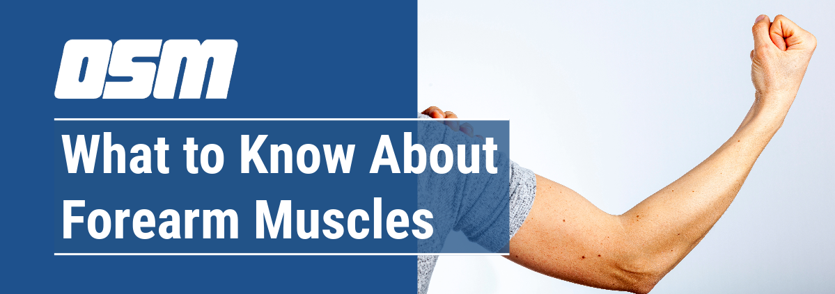
Your bicep is the muscle in the front of your upper arm. It helps you bend your elbow and twist your forearm.

Three tendons attach your bicep to bone:

  • The long head tendon attaches your bicep to the top of your shoulder socket.
  • The short head tendon attaches your bicep to a bump on your shoulder blade called the coracoid process.
  • A third tendon attaches your bicep to your radius, which is one of the bones in your forearms.

When you have a torn bicep, one of these tendons is damaged or detaches from the bone. Any of these three bicep tendons can tear.

Types of bicep tendon tear injuries

There are three types of bicep tendon tear injuries, categorized by their location and severity. Tears can also be partial (in which a tendon is damaged) or complete (in which the tendon completely detaches from the bone).

The three types of bicep tendon tear injuries are:

Proximal biceps tendon tear at shoulder

This injury occurs when one of the tendons that attaches the bicep to the shoulder tears. The long head tendon is more likely to tear than the short head tendon. This type of tear often starts as normal tendon fraying, but can also tear if you get injured.

It’s likely that only one part of the tendon will tear in this injury. This means that you can usually continue to use your arm. However, a bicep tendon tear at the shoulder may damage other parts of the shoulder at the same time.

Distal biceps tendonitis and tear at the elbow

A bicep tendon tear at the elbow usually happens when the elbow is pushed straight against a heavy weight. This stress can tear the tendon from the bone, and usually causes a complete tear.

When you tear your bicep tendon at the elbow, your other arm muscles will compensate, so you’ll still have full range of motion. However, your arm will most likely lose strength if the tendon is not repaired. Bicep tendon tears at the elbow are not common. They happen to approximately 3 to 5 people per 100,000 per year. They’re also less common in women. Distal biceps tendonitis is inflammation in the biceps tendon near the elbow. It’s usually caused by normal wear and tear but repetitive motion can make it worse.

Tendonitis (microtears from use)

Tendonitis is the inflammation or irritation of the long head of the bicep tendon. This can cause microtears. As with distal biceps tendonitis, tendonitis of the long head of the biceps tendon is usually due to normal wear and tear, but can also be made worse by repetitive motion. It often happens with other shoulder problems, such as arthritisshoulder impingement, and chronic shoulder dislocation.

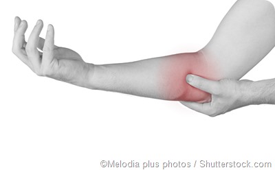
Torn bicep tendon symptoms

Symptoms of a torn bicep tendon include:

  • a “pop” or tearing sensation when the injury happens
  • warmth around the injury
  • swelling
  • bruising
  • pain or ache at the injury site, and throughout your arm (usually severe at first, and may get better over a few weeks)
  • arm weakness
  • difficulty turning your palm
  • fatigue or increased pain in your arm when you do repetitive activity
  • bulge in your upper arm, because the bicep is no longer being held in place (you might also see a gap or indentation in front of your elbow)

Causes of a torn bicep tendon

The two main causes of a torn bicep tendon are injury and overuse. Injuries might be caused by lifting something heavy or falling on your arm. Most tears of the elbow bicep tendon happen because of an injury.

Overuse can cause the tendons to wear down or fray over time. This happens naturally as you age. It may also be made worse by repetitive motion, and is common in people who participate in sports such as weightlifting, tennis, or swimming.

Diagnosing a torn bicep tendon

To diagnose a torn bicep tendon, a doctor will first take a medical history. They’ll ask about your symptoms, whether you had any recent injuries, and when the pain began.

Then they’ll do a physical exam to test your range of motion and strength. During these tests, they’ll see if you have pain or difficulty with certain movements, especially rotations. They’ll also look at your arm for swelling, bruising, or bulging.

A history and physical exam are often enough to diagnose a bicep tendon tear. However, your doctor might also do an X-ray to help rule out any bone injuries, or an MRI to see if the tear is partial or complete.

Torn bicep treatment

Treatment for a torn bicep will mostly depend on how severe the tear is, as well as your overall bicep function and whether you damaged any other body part, such as your rotator cuff. Potential treatments include:

Rest

Taking time off from exercising, lifting, or holding anything heavy — and using your arm as little as possible — can help you recover, especially from overuse injuries. Be sure to avoid any activity that causes pain, even if it doesn’t seem strenuous.

NSAIDs

Nonsteroidal anti-inflammatory drugs (NSAIDs) are over-the-counter medications that help reduce inflammation. They can help reduce the inflammation (the hallmark of tendonitis), as well as help reduce swelling from bicep tears. They can also help reduce the pain you might have from any bicep tendon injuries.
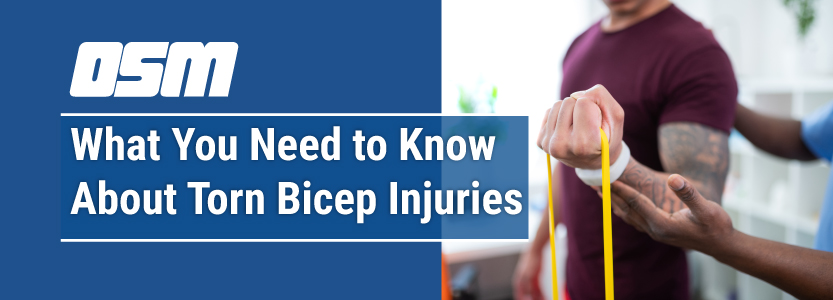
Physical therapy

Physical therapy can help you regain strength and range of motion after a bicep tendon injury. A physical therapist will take you through a series of motions designed to help heal your injury and relieve pain.

A physical therapist or your doctor might also give you exercises to do at home when you’re healed enough to do so. These might include exercises to flex and extend your arm, arm rotations, and strength-building exercises like bicep curls.

Torn bicep surgery

If none of the measures above help your bicep injury heal, or if more than half the tendon is torn, your doctor might recommend surgery to repair the bicep tendon.

Many doctors will recommend surgery as a first-line treatment for bicep tendon tears at the elbow, although surgery can also be done later if other treatments don’t restore range of motion and strength.

Surgery is used to reattach the tendon to the bone. Complications of surgery are rare, but may include arm numbness or weakness. In some people, the tendon can tear again.

Torn bicep tendon recovery time

Recovery time depends on the severity of the bicep tendon tear, as well as type of treatment. Even mild injuries can take at least two months to heal. It often takes four to five months before you can start returning to normal activities.

After surgery, you’ll probably need to wear a sling or otherwise immobilize your arm such as in a splint or cast for four to six weeks. You’ll then have to do physical therapy and exercises to help strengthen your arm and improve range of motion.

Complete recovery from surgery can take up to a year, although most people recover much of their range of motion and strength in four to six months.

Takeaway

Bicep tendon tears can be serious, but many respond to nonsurgical treatment, such as rest and physical therapy. If you think you might have injured your bicep tendon, see a doctor as soon as possible. Getting a diagnosis and treatment early can help you recover more fully.


The Orthopedic & Sports Medicine Center of Oregon is an award-winning, board-certified orthopedic group located in downtown Portland Oregon. We utilize both surgical and nonsurgical means to treat musculoskeletal trauma, spine diseases, sports injuries, degenerative diseases, infections, tumors and congenital disorders.

Our mission is to return our patients back to pain-free mobility and full strength as quickly and painlessly as possible using both surgical and non-surgical orthopedic procedures.

Our expert physicians provide leading-edge, comprehensive care in the diagnosis and treatment of orthopedic conditions, including total joint replacement and sports medicine. We apply the latest state-of-the-art techniques in order to return our patients to their active lifestyle.

If you’re looking for compassionate, expert orthopedic surgeons in Portland Oregon, contact OSM today.

Phone:
503-224-8399

Address
1515 NW 18th Ave, 3rd Floor
Portland, OR 97209

Hours
Monday–Friday
8:00am – 4:30pm

How Do You Know if You Fractured Your Forearm?

Article featured on Raleigh Orthopaedic

The forearm is made up of two bones in your lower arm, the radius and ulna. A fracture in the forearm can occur near the wrist, in the middle of the forearm or near the elbow. The forearm motion allows us to rotate our palms up or down. A broken forearm can affect your ability to rotate your arm and even bend or straighten the wrist and elbow.

A bone may be completely fractured or partially fractured in any number of ways (crosswise, lengthwise, in multiple pieces). If the bone breaks in such a way that bone fragments stick out through the skin, or a wound penetrates down to the broken bone, the fracture is called an “open” fracture and requires immediate attention. Call 911 or go to your nearest emergency room.

Cause of a fractured forearm

  • A direct blow to the forearm
  • Fall on an outstretched arm

What are the symptoms of a broken forearm?

  • Immediate pain in the lower arm
  • Obvious deformity
  • Swelling
  • Bruising
  • Inability to rotate the arm

To determine whether you have broken forearm, your physician will ask you for a complete medical history and conduct a physical examination. An X-ray can confirm the diagnosis.

How do you treat a fractured forearm?

In an emergency room or urgent care, forearm fracture treatment begins with stabilizing the bones and temporarily realigning them with a cast or splint.

Nonsurgical treatment for a broken forearm

If the broken bone is in a good position or can be put back into good alignment, a cast can be used to keep the forearm steady until it heals. An orthopedic physician will monitor the healing of the fracture with frequent clinic visits and X-rays.

Broken forearm surgery

If the broken bone is out of place and the alignment cannot be corrected, surgery may be required. Surgery is also necessary for open fractures where the bones have broken through the skin. This type of forearm surgery depends on where the break in the bones occurred and the severity of the break. Your surgeon will determine the best surgery for your injury.

Rehabilitation

Recovery for a broken forearm depends on the severity of the injury. Forearm bones typically take three to six months to fully heal. The more severe your injury, the longer the recovery may be.

Rehabilitation usually begins are a few weeks of healing. Sometimes you may still use a cast or brace during the beginning stages of physical therapy. Physical therapy will focus on strength and range of motion exercises.


The Orthopedic & Sports Medicine Center of Oregon is an award-winning, board-certified orthopedic group located in downtown Portland Oregon. We utilize both surgical and nonsurgical means to treat musculoskeletal trauma, spine diseases, sports injuries, degenerative diseases, infections, tumors and congenital disorders.

Our mission is to return our patients back to pain-free mobility and full strength as quickly and painlessly as possible using both surgical and non-surgical orthopedic procedures.

Our expert physicians provide leading-edge, comprehensive care in the diagnosis and treatment of orthopedic conditions, including total joint replacement and sports medicine. We apply the latest state-of-the-art techniques in order to return our patients to their active lifestyle.

If you’re looking for compassionate, expert orthopedic surgeons in Portland Oregon, contact OSM today.

Phone:
503-224-8399

Address
1515 NW 18th Ave, 3rd Floor
Portland, OR 97209

Hours
Monday–Friday
8:00am – 4:30pm

Cubital and Radial Tunnel Syndrome: What Does That Mean?

Article featured on WebMD

Cubital tunnel syndrome and radial tunnel syndrome aren’t as familiar as their better-known relative — carpal tunnel syndrome — but they also can cause severe pain, numbness, tingling, and muscle weakness in the hands and arms.The common cause of all these nerve compression syndromes is increased pressure — usually from bone or connective tissue — on a nerve in the wrist, arm, or elbow. In most cases, cubital tunnel syndrome and radial tunnel syndrome can be managed with conservative treatments. But more severe cases may require surgery to reduce pressure on the affected nerve.

Cubital Tunnel Syndrome: Causes and Symptoms

Cubital tunnel syndrome — also known as ulnar neuropathy — is caused by increased pressure on the ulnar nerve, which passes close to the skin’s surface in the area of the elbow commonly known as the “funny bone.” You’re more likely to develop cubital tunnel syndrome if you:

  • Repeatedly lean on your elbow, especially on a hard surface
  • Bend your elbow for sustained periods, such as while talking on a cell phone or sleeping with your hand crooked under your pillow
Sometimes, cubital tunnel syndrome results from abnormal bone growth in the elbow or from intense physical activity that increases pressure on the ulnar nerve. Baseball pitchers, for example, have an increased risk of cubital tunnel syndrome, because the twisting motion required to throw a slider can damage delicate ligaments in the elbow.Early symptoms of cubital tunnel syndrome include:

  • Pain and numbness in the elbow
  • Tingling, especially in the ring and little fingers

More severe symptoms of cubital tunnel syndrome include:

  • Weakness affecting the ring and little fingers
  • Decreased ability to pinch the thumb and little finger
  • Decreased overall hand grip
  • Muscle wasting in the hand
  • Claw-like deformity of the hand

If you have any of these symptoms, your doctor may be able to diagnose cubital tunnel syndrome by physical examination alone. They also may order a nerve conduction study and a test called electromyography. Electromyography is a procedure in which electrodes placed into muscles and on the skin measure the health of muscles and the nerve cells that control them, to confirm the diagnosis, identify the area of nerve damage, and determine the severity of the condition.

Radial Tunnel Syndrome: Causes and Symptoms

Radial tunnel syndrome is caused by increased pressure on the radial nerve, which runs by the bones and muscles of the forearm and elbow. Causes include:

  • Injury
  • Noncancerous fatty tumors (lipomas)
  • Bone tumors
  • Inflammation of surrounding tissue

Symptoms of radial tunnel syndrome include:

  • Cutting, piercing, or stabbing pain at the top of the forearm or back of the hand, especially when you try to straighten your wrist and fingers.

In contrast to cubital tunnel syndrome and carpal tunnel syndrome, radial tunnel syndrome rarely causes numbness or tingling, because the radial nerve principally affects the muscles.

Just as with cubital tunnel syndrome, if you have any of these symptoms, your doctor may be able to diagnose radial tunnel syndrome by physical examination alone. They also may order electromyography to confirm the diagnosis, identify the area of nerve damage, and stage the severity of the condition.

Treatments for Cubital Tunnel Syndrome and Radial Tunnel Syndrome

Cubital tunnel syndrome often can be managed conservatively, especially if electromyography reveals that there is minimal pressure on the ulnar nerve.

Mild cases of cubital tunnel syndrome often respond to physical therapies such as:

  • Avoidance of undue pressure on the elbow during daily activities
  • Wearing a protective elbow pad over the “funny bone” during daily activities
  • Wearing a splint during sleep to prevent over-bending of the elbow

In cases where splinting doesn’t help or nerve compression is more severe, about 85% of patients respond to some form of surgery to release pressure on the ulnar nerve. These include surgeries that:

  • Result in simple decompression of the ulnar nerve
  • Shift the nerve to the front of the elbow
  • Move the nerve under a layer of fat, under the muscle, or within the muscle
  • Trim the bump of the inner portion of the elbow — the medial epicondyle — under which the ulnar nerve passes

If you undergo surgery for cubital tunnel syndrome, recovery may involve restrictions on lifting and elbow movement, and rehabilitation therapy. Although numbness and tingling may or may not quickly improve, recovery of hand and wrist strength may take several months.

Conservative treatments for radial tunnel syndrome include medications such as nonsteroidal anti-inflammatory drugs to reduce soft tissue swelling, corticosteroid injections to relieve inflammation and pressure on the radial nerve, and wrist and/or elbow splints to reduce irritation of the radial nerve.Some patients also may benefit from ergonomic education to reduce the effects of repetitive stress, nerve-gliding exercises, stretching/strengthening exercises, and other interventions such as heat, cold, and ultrasound.If these conservative measures fail to provide relief after three months, your doctor may consider surgery to reduce pressure on the radial nerve. Surgery is often recommended in severe cases, particularly those in which the wrist becomes weak or droopy or it becomes difficult to extend the fingers.

The Orthopedic & Sports Medicine Center of Oregon is an award-winning, board-certified orthopedic group located in downtown Portland Oregon. We utilize both surgical and nonsurgical means to treat musculoskeletal trauma, spine diseases, sports injuries, degenerative diseases, infections, tumors and congenital disorders.

Our mission is to return our patients back to pain-free mobility and full strength as quickly and painlessly as possible using both surgical and non-surgical orthopedic procedures.

Our expert physicians provide leading-edge, comprehensive care in the diagnosis and treatment of orthopedic conditions, including total joint replacement and sports medicine. We apply the latest state-of-the-art techniques in order to return our patients to their active lifestyle.

If you’re looking for compassionate, expert orthopedic surgeons in Portland Oregon, contact OSM today.

Phone:
503-224-8399

Address
1515 NW 18th Ave, 3rd Floor
Portland, OR 97209

Hours
Monday–Friday
8:00am – 4:30pm

Causes of Elbow or Arm Pain

Article features on News Medical Life Sciences

There are various possible causes of elbow pain or arm pain. These include injury to the arm, compression of the nerves that serve the arm, arthritis, and various other health conditions.

elbow pain

Injury

A sprain can occur when a movement of the arm has caused damage to the connective tissues in the area. This can present as arm or elbow pain. This damage is usually acute and temporary, and the pain will improve with time as the tissues are repaired.

A fracture or dislocation of a bone in the arm or elbow may also be responsible for causing elbow pain. Most patients are able to pinpoint the cause of the pain or trace it to a certain incident that caused the damage.

Repetitive strain injury (RSI) can also cause elbow pain. It is due to the performance of repetitive tasks that involve movements of the elbows. As such, it commonly affects office workers who use computers frequently throughout the day, and other individuals whose job comprises repetitive manual work.

Tennis elbow or golfer’s elbow can cause elbow pain, and occurs due to the overuse of the muscles and tendons in the joint. It is so named because it is most common in individuals who plan tennis or golf, due to the arm movements that are involved in these sports.

Nerve compression

The joints and bones of an individual show wear and tear as part of the natural aging process. This can lead to the squashing or trapping of nerves, which in turn can cause pain in various areas of the body. In this case, the nerves around the elbow may be pinched, causing pain. Other sensations, such as numbness or tingling, may also be experienced.

cubital tunnel syndrome

Cervical spondylosis is a condition that involves compression of the spinal nerves in the neck region. Some of these nerves are responsible for the sensory messages of pain carried to and from the arm. Damage to them can hence lead to arm pain. Sometimes the compressed nerve may be located in the arm.

A cervical rib is a musculoskeletal abnormality that involves the presence of an additional rib above the normal first rib. This extra bone can interfere with the free passage of nerves in the region, which may lead to elbow pain.

Arthritis

Osteoarthritis or rheumatoid arthritis that affects the elbow joint can lead to stiffness, pain and inflammation of the elbow and arm.

Osteoarthritis involves damage to the cartilage at the ends of the bones that help to prevent friction between the bones that take part in the joint, often due to overuse of or injury to the joint. This leads to rubbing of the bones against each other, which provokes the symptoms of arthritis.

Rheumatoid arthritis involves an autoimmune response of the body that leads to damage to the synovial membrane that lines the joints in the body, including the elbow. In this condition, both elbows are usually affected.

Other health conditions that cause elbow pain

Angina is a health condition that involves reduced blood flow to the heart muscles as a result of narrowing of the blood vessels. Individuals with angina may experience a dull, throbbing pain in the chest, neck, and left arm as a symptom of the condition, particularly during physical activity or when they undergo stress.

Gout is another health condition characterized by the formation of sharp crystals at various locations around the body, including inside the elbow joints. This is due to a high concentration of uric acid in the body. This can cause severe elbow pain during a gout attack.

Olecranon bursitis is the build up of fluid inside the fold of smooth synovial membrane that cushions the olecranon (the bony tip of the elbow joint). This can cause pain and inflammation.


The Orthopedic & Sports Medicine Center of Oregon is an award-winning, board-certified orthopedic group located in downtown Portland Oregon. We utilize both surgical and nonsurgical means to treat musculoskeletal trauma, spine diseases, sports injuries, degenerative diseases, infections, tumors and congenital disorders.

Our mission is to return our patients back to pain-free mobility and full strength as quickly and painlessly as possible using both surgical and non-surgical orthopedic procedures.

Our expert physicians provide leading-edge, comprehensive care in the diagnosis and treatment of orthopedic conditions, including total joint replacement and sports medicine. We apply the latest state-of-the-art techniques in order to return our patients to their active lifestyle.

If you’re looking for compassionate, expert orthopedic surgeons in Portland Oregon, contact OSM today.

Phone:
503-224-8399

Address
1515 NW 18th Ave, 3rd Floor
Portland, OR 97209

Hours
Monday–Friday
8:00am – 4:30pm

Care of Casts and Splints

Care of Casts and Splints

Article Featured on AAOS

Casts and splints support and protect injured bones and soft tissue. When you break a bone, your doctor will put the pieces back together in the right position. Casts and splints hold the bones in place while they heal. They also reduce pain, swelling, and muscle spasm.

Read more